INTRODUCTION
Spinal cord infarction is a rare condition and represents a diagnostic challenge for emergency physicians. Since the initial manifestation of spinal cord infarction is non-specific, it is easily misdiagnosed. Pseudoangina, mimicking the pain of coronary ischemia, is a non-cardiogenic chest pain. We present the case of a 77-year-old man who initially presented to the emergency department with chest and back pain only, and was diagnosed as having a spinal cord infarction.
CASE REPORT
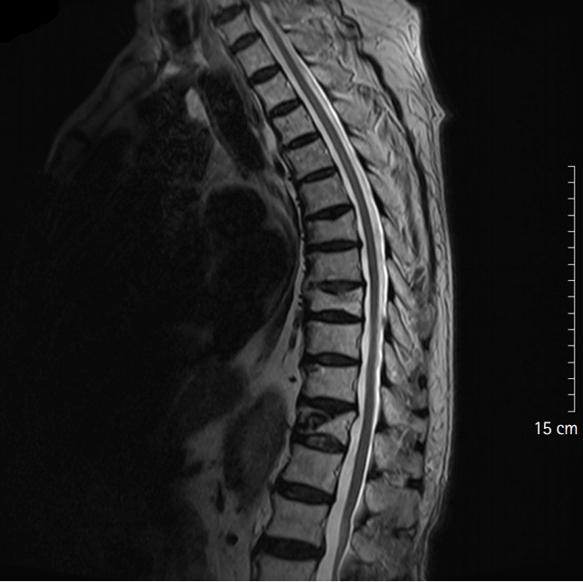
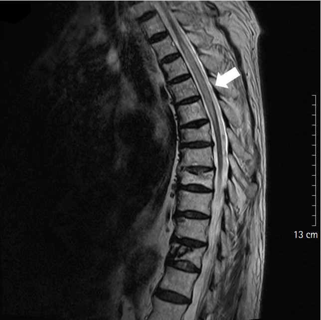
A 77-year-old man visited an emergency department for chest pain that developed 1 hour before the hospital visit. The pain began when he washed his face after getting out of bed in the morning. The patient felt a tightening pain in the chest, through the back, and in the area between both the scapulae. The patient was taking aspirin 100 mg, clopidogrel 75 mg, and metformin 500 mg for hypertension and diabetes. He visited the hospital one year prior for a similar pain and underwent a stent insertion in the left anterior descending coronary artery for ST elevation myocardial infarction. When the patient visited the hospital this time, the pain was rated as an 8 out of 10 and continued even after sublingual administration of the patients own nitroglycerin. Vital signs were as follows: blood pressure, 166/88 mmHg; pulse rate, 72 beats/min; respiratory rate, 15 breaths/min; and body temperature, 36.7┬░C. Sinus rhythm and first-degree atrioventricular block were observed on electrocardiography. Bedside echocardiography showed similar results with the previous one, except for a decrease in ejection fraction to 38%. Laboratory data including hemoglobin, platelet count, white blood cell, blood chemistry, and cardiac enzyme test as well as urinalysis were normal. Thirty minutes after the hospital admission, the chest pain was relieved; however, the pain through the scapula remained. Therefore, aortic dissection was suspected and an aortic dissection computed tomography was obtained. The computed tomography showed multiple dissections at an infrarenal level. Since the patient had slipped and fallen 3 days prior, spinal radiography was performed and revealed an acute compression fracture at T8. Four hours later, he complained of numbness in the entire left leg below the knee. The numbness alternately occurred in the left and right leg during a neurological examination but the motor activity was measured as grade 5. Spinal magnetic resonance imaging (MRI) was obtained to check if a spinal cord injury was caused by the compression fracture, but there was no definite signal change in the spinal cord (Fig. 1). Six hours after the visit, the upper back pain increased in severity to the point that the patient was unable to lift his knee up, and the motor activity of the lower limb decreased to grade 3. A spinal cord infarction was suspected and the patient was admitted to the department of neurology. Nine hours after the hospital visit, a sudden sensory changeŌĆōhypoesthesia (3/10)ŌĆōoccurred, loss of sensation progressed up to the T4 dermatome, the strength in both lower extremities deteriorated to grade 0, and a decrease in anal tone and deep tendon reflex was observed. The patient was numb to pain and temperature stimuli, but vibratory sensation was intact. Steroid pulse therapy (methylprednisolone 1 g/day) was initiated. Seventy-two hours after the hospital admission, no change in the neurological symptoms was observed. A follow-up spinal MRI showed a signal change with a diffuse pattern within the spinal cord at T2-T6 (T2 hyperintensity, T1 hypointensity without definite enhancement), and spinal cord swelling was observed (Fig. 2). Steroid pulse therapy was continued for 5 days. After a month of hospitalization, touch sensation in his right big toe had improved slightly, but the patient was still undergoing rehabilitation treatment while showing no improvement in the other neurological symptoms.
DISCUSSION
Non-traumatic chest pain is one of the most common complaints in the emergency department. The incidence of myocardial infarction with chest pain has been estimated at about 10%, whereas other life-threatening conditions include pulmonary embolism, aortic dissection, and spontaneous tension pneumothorax [1]. Less emergent, and common conditions including gastroesophageal reflux, costochondritis, pleuritic, and other musculoskeletal conditions also need to be included in the differential diagnosis of non-traumatic chest pain [2]. Pseudoangina, mimicking the pain of coronary ischemia, is a cause of non-cardiogenic chest pain [3]. Sensory neurons in the upper thoracic spinal cord transmit the pain of myocardial ischemia from the cardiac plexus [4].
Spinal cord infarction is rare, and its etiologies are extensive including atheroembolic disease, complications from aortic aneurysm repair, aortic dissection, vasculitis, infection, prolonged hypotension, and trauma [5].
The initial manifestations of spinal cord infarction are non-specific and it is easily misdiagnosed. Spinal cord infarction is usually marked by an acute onset, often heralded by sudden and severe back pain, which may radiate caudad. This is associated with bilateral weakness, paresthesias, and sensory loss. Loss of sphincter control with hesitancy and inability to void or defecate can occur. Early symptoms of high-level thoracic spinal cord infarction include upper back pain and chest pain, which can be mistaken for emergent cardiopulmonary conditions including acute myocardial infarction, pulmonary embolism, aortic dissection, and pneumothorax [1]. In our case, the initial presentation with acute chest pain and upper back pain and the patientŌĆÖs history of myocardial infarction misled the attending emergency physician to focus on an acute myocardial infarction and aortic dissection. Neurological deficits may occur without pain, but most (>80%) spinal infarcts are painful [6]. This is an interesting and unexplained difference from cerebral infarction, which is usually not painful.
MRI is the most sensitive and reliable tool to detect spinal cord infarction. It can show signal changes and swelling in the cord within a few hours of onset, but it has some important limitations [7-9]. An MRI finding early in the course of spinal cord infarction may be normal, even in the presence of significant neurological deficits [5]. In our case, MRI findings showed a signal change 3 days after the symptoms developed.
Currently, there is no treatment that is known to facilitate an improvement in patients who have spinal cord infarction. Treatment focuses on risk factors and rehabilitation. Aspirin therapy is often initiated and no clinical trials support the use of thrombolytics. Some studies showed that corticosteroid therapy initiated within a short time after spinal cord infarction had protective effects on cell function and reduced oxidative stress following ischemic injury [10]. Treatment with rehabilitative therapy remains supportive. Prognosis is predicted based on the MRI appearance of the spinal cord and the clinical presentation [5,11]. Recovery following spinal cord infarction is diverse. Twenty-two percent of spinal cord infarction patients die during their initial hospitalization [5]. Some studies have shown that only 18% to 41% of survivors can ambulate independently at hospital discharge, while 20% to 57% will remain wheelchair-dependent. Functional recovery is best predicted by the initial degree of motor dysfunction [5,12].
In the patient with acute chest pain combined with neurologic symptoms, one should consider not only the possibility of ischemic cardiac disease but also evaluate any neurological manifestations. Emergency department physicians must remember the value of serial physical examinations and ensure rapid access to MRI to rule out spinal lesions.