Descending necrotizing mediastinitis after a trigger point injection
Article information
Abstract
Descending necrotizing mediastinitis (DNM) is a rare form of mediastinal infection. Most cases are associated with esophageal rupture. DNM after a trigger point injection in the upper trapezius has not been described previously. We present a case of DNM after a trigger point injection in the upper trapezius. A 70-year-old man visited the emergency department with chest discomfort and fever after a trigger point injection in the left upper trapezius. Chest computed tomography showed evidence of DNM, and antibiotic therapy was immediately administered intravenously. Because of the risk of sudden death, poor prognosis due to underlying disease, and his age, he declined surgical treatment and died of septic shock. Although trigger point injections are generally considered safe, caution should be used in patients with an underlying disease or in the elderly. Early diagnosis, broad-spectrum antibiotics, and aggressive surgical management are essential to improve the prognosis.
INTRODUCTION
Mediastinitis is an inflammation of the connective tissues that involves mediastinal structures and fills the interpleural space [1]. Descending necrotizing mediastinitis (DNM) is a serious, life-threatening complication of mediastinal infection that can originate in the esophagus (through operation or rupture), or from oropharyngeal infection or cervical trauma. DNM is caused by the downward spread of a head and neck infection to the mediastinum through connecting deep and superficial cervical fascial planes [2]. The risk factors for DNM include intravenous drug use, alcoholism, old age, and chronic debilitating comorbidities (diabetes mellitus, underlying malignancy, liver cirrhosis, and renal failure) [3]. DNM can be secondary to infectious or non-infectious causes and, depending on the etiology, can be acute or chronic. Diagnostic criteria for DNM were proposed by Estrera et al. [4] in 1983: clinical manifestations of severe infection; characteristic radiographic features of mediastinitis; documentation of a necrotizing mediastinal infection during an operation, at autopsy, or both; and establishment of the relationship between DNM and an oropharyngeal or cervical infection. Despite advanced diagnostic and surgical techniques, DNM with sepsis reportedly has high rates of mortality, primarily because of delayed diagnosis [5]. However, the diagnosis of DNM can be difficult, owing to the vagueness of the symptoms [6]. DNM after a trigger point injection (TPI) has not been described previously; here, we present a case of DNM after TPI in the left upper trapezius.
CASE REPORT
A 70-year-old Asian man with diabetes mellitus treated with oral hypoglycemic agents and a history of myocardial infarction managed with percutaneous coronary intervention and antiplatelet drugs visited the emergency department (ED) of our hospital complaining of 7 days of progressive chest discomfort, fever, and chills. Ten days prior, he visited a local clinic with left upper back and shoulder pain. He was diagnosed with myofascial pain syndrome and received a TPI with lidocaine using a 21-gauge, 2.0-inch needle in the left upper trapezius. Three days after the injection, he complained of substernal chest pain of a squeezing nature and chills, and visited the ED of another hospital. Coronary angiography at that hospital was normal. His symptoms progressively worsened despite administration of analgesics, and he was transferred to our hospital.
He was an active smoker, with a cumulative exposure of 35 pack-years, and a heavy drinker, consuming 1 bottle of alcohol daily. He did not have clinical evidence of a recent head and neck infection or trauma. He was 163 cm tall and weighed 60.3 kg (body mass index, 22.7 kg/m2). In the ED, his blood pressure was 120/78 mmHg, heart rate was 110 beats/min, oxygen saturation was 97%, and axillary temperature was 38.2°C. A physical examination revealed tenderness and redness from the left side of the upper sternum to the same side of the shoulder. The other examination results were normal.
Laboratory data were significant for a white blood cell count of 19.9×10⁹/L, with 86.7% neutrophils. Both C-reactive protein and erythrocyte sedimentation rate were elevated, at 26.5 mg/L (reference, <0.5 mg/dL) and 80 mm/hr (0–10 mm/hr), respectively. Arterial blood gas analysis showed pH 7.48, PCO2 28 mmHg, PO2 115.3 mmHg, and HCO3 20.9 mmol/L. Serum glucose was 249 mg/dL and a urine test strip was 4+ for glucose. Other laboratory test results were normal, including plasma levels of electrolytes, troponin, and creatinine kinase-MB. Aerobic and anaerobic blood cultures were drawn before empirical antibiotic treatment.
Chest radiography showed upper mediastinal widening, protrusion of the aortic arch, and consolidation in the left upper lobe (Fig. 1). Contrast-enhanced chest computed tomography (CT) revealed a 3.8×3-cm outpouching low-density lesion with an infiltrative margin arising from the aortic arch, diffuse mild wall-thickening in the aorta, and an incidental 6.8-cm heterogeneous enhancing mass in the right hepatic lobe (Fig. 2). Magnetic resonance imaging of the liver suggested hepatocellular carcinoma in the S7 segment.

Chest radiography. Consolidation is seen in the left upper lobe of the lung.
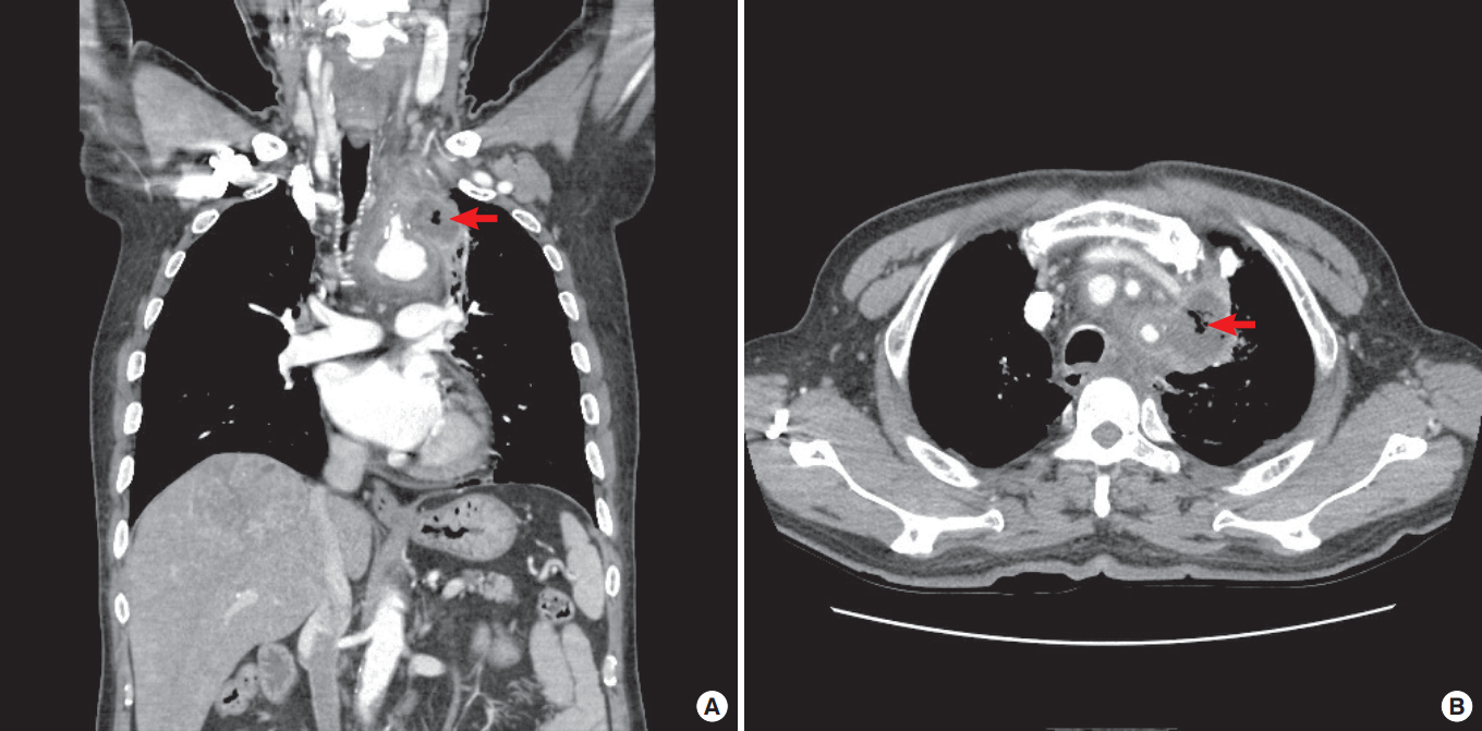
(A) Contrast-enhanced chest computed tomography (CT, coronal plane). (B) Contrast-enhanced chest CT (axial plane). Chest CT showing well-defined, lobulated fluid collections with gas bubbles in the aortic arch (arrow).
He was treated immediately with intravenous ceftriaxone and metronidazole, and the initial blood culture revealed Staphylococcus aureus. Based on clinical and radiological features, he was diagnosed with DNM after TPI.
Fever persisted following the ED visit, and both C-reactive protein and erythrocyte sedimentation rate remained elevated, at 23.6 mg/L and 37 mm/hr, respectively. Antibiotics were changed on hospital day 3 to piperacillin/tazobactam. However, intermittent fever persisted until hospital day 5, and finally subsided when the antibiotics were changed to meropenem and vancomycin.
On hospital day 9, he had hemoptysis. Follow-up CT showed increased mediastinal infiltration and a low-density lesion with an air bubble and a penetrating atherosclerotic ulcer at the aortic arch (Fig. 3). Based on the CT, we explained the process of surgical drainage. However, he and his family refused aggressive management such as surgery, because of the risk of sudden death during the operation, poor prognosis of underlying hepatocellular carcinoma, and his age. Despite antibiotic therapy, his general condition worsened, and he authorized a do-not-resuscitate order. On hospital day 30, he died because of septic shock.
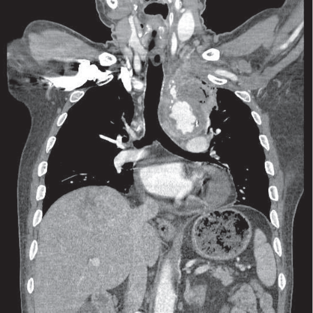
Follow-up chest computed tomography after 8 days. Increased mediastinal infiltration and a low-density lesion with an air bubble are visible.
DISCUSSION
The majority of DNM cases are associated with cardiovascular operations, esophageal perforation, tracheobronchial perforation, mediastinal extension of pulmonary infections, or mediastinal extension of head and neck infections. However, to the best of our knowledge, DNM following TPI in the upper trapezius has not been described.
Trigger points are focal hyperirritable areas within a taut band of skeletal muscle that are painful on precise compression. Trigger points can be very painful and disabling and even cause weakness. The rhomboid and trapezius muscles, located in the upper back and shoulder areas, are common sites of trigger points [7]. A TPI is one of the most effective therapeutic approaches, and is generally considered very safe. The most common complication is infection, including osteomyelitis, epidural abscess, and necrotizing fasciitis. Noninfectious complications include peripheral nerve injuries, pneumothorax, air embolism, and pain or swelling at the site of the injection [8].
The diagnosis of DNM is often difficult due to nonspecific and vague early symptoms, which include chills, fever, tachycardia, dyspnea, and a nonproductive cough [2,3,5]. A CT scan can immediately confirm mediastinitis with high accuracy, showing soft tissue infiltration with loss of the normal fat planes or collection of fluid with or without the presence of gas bubbles. The use of contrast-enhanced chest CT is the diagnostic method of choice for patients in whom DNM is suspected [2-4].
Treatment for DNM includes broad-spectrum antibiotics and early aggressive surgical intervention. Intravenous broad-spectrum antibiotic therapy is not effective without adequate surgical drainage of cervical and mediastinal collections and extensive debridement and excision of necrotic tissue. In reported cases of DNM, successful surgical management involves a combination of cervical and mediastinal drainage, with or without open thoracotomy [1,3,4,9,10]. More recently, less invasive approaches that successfully employ thoracoscopic or mediastinoscopic drainage have been reported [11,12].
The present case highlights the importance of collecting the patient history, physical examination, and radiographic findings to differentiate between DNM and other causes that present similar symptoms, including chest pain. Although TPI is generally safe, a complaint of chest discomfort or pain, fever, cough, and dyspnea following a TPI in the neck area should prompt consideration of DNM. A high index of suspicion and appropriate investigations are important, as a favorable outcome depends on early diagnosis and surgical intervention. In summary, early diagnosis, aggressive medical and surgical interventions, and close surveillance with serial CT are crucial for reducing the mortality associated with DNM.
Notes
No potential conflict of interest relevant to this article was reported.
References
Article information Continued
Notes
Capsule Summary
What is already known
Trigger point injections are generally considered safe. Descending necrotizing mediastinitis is a serious, life-threatening infection that can originate in the esophagus (through operation or rupture), or from oropharyngeal infection or cervical trauma.
What is new in the current study
Although trigger point injections are generally safe, a complaint of chest discomfort or pain, fever, cough, and dyspnea following a trigger point injection in the neck area should prompt consideration of descending necrotizing mediastinitis.