INTRODUCTION
Clavicle fractures are less common in infants than in older children [1]. It is well known that most often, these fractures are caused by falls and are treated non-operatively [2]. The frequency of skull fractures in infants and the proximity of the clavicle to the skull could predispose the clavicle to fractures, and the anterior-posterior view of the skull (skull AP) on an X-ray series includes the clavicle in infants. However, if physicians order skull APs to assess skull fractures, they may miss unexpected clavicle fractures. To our knowledge, no such cases have been reported, and many clavicle fractures in infants demonstrate a favorable prognosis. However, the regular use of the skull AP could result in unintended neglect of fractures, with subsequent complications, and even medico-legal problems. The objectives of this study were to determine how often the clavicle and associated fractures are visible but missed on the skull AP and which factors are associated with a missed diagnosis.
METHODS
Study design and setting
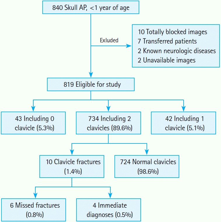
This was a retrospective observational descriptive study. We identified patients who presented between April 1999 and July 2012 to the pediatric emergency department (PED) of our hospital and were evaluated using the skull AP for any injuries. Our hospital is an urban, academic hospital with approximately 3,000 beds, and has a PED with about 40,000 visits annually. We included patients aged 1 year or younger who were evaluated using the skull AP, but not necessarily an entire skull X-ray series, as part of any injury assessment. We excluded patients for the following reasons: entire clavicle not visible on X-ray, ŌĆ£including 0-1 clavicleŌĆØ; known neurologic disorders; images taken at other institutions; images unreadable due to poor quality, guardianŌĆÖs hands or soft tissue density; images unavailable because of technical problems (Fig. 1).
Data extraction
Medical records were obtained by searching the electronic medical records for the key word ŌĆ£skull X-ray.ŌĆØ Hospital policy requires that all children with a head injury receive a skull X-ray.
Initially, the percentage of visible clavicles on the skull AP was determined and recorded as ŌĆ£including 0-2 claviclesŌĆØ (Fig. 1). The clavicle was defined as ŌĆ£visibleŌĆØ if 50% was recognizable on each side. Among patients who were classified as ŌĆ£including 2 clavicles,ŌĆØ we identified all clavicle fractures. Clavicle fractures were defined as bony discontinuity of the clavicle demonstrated on skull AP. In cases of unavailable comments on formal reports, 2 emergency medicine board-certified physicians who were not blinded to study objectives discussed the findings and made the final decisions. The skull AP was used as the reference standard because of its feasibility for diagnosis and its frequent use. Accordingly, clavicle fractures that were unrecognizable on the skull AP but recognizable on other images (e.g., lateral or TowneŌĆÖs view of the skull, clavicle X-ray) were not used for diagnosis.
We investigated patientsŌĆÖ age, sex and injury mechanisms. We also assessed the accuracy of the diagnosed clavicle fractures. Only cases having descriptions of the clavicle included on sameday charts were considered accurate. In our clinical experience, skull AP interpretation can be hindered by the guardianŌĆÖs hands holding the patientŌĆÖs chin. Therefore, we determined whether or not the guardianŌĆÖs hands were obstacles to interpretation. In addition, we investigated if skull fractures, as the main indication for skull AP, were an obstacle to interpretation.
Statistical analysis
Demographic data and clinical manifestations were investigated. We also determined the associations between clinical findings and missed clavicle fractures. Categorical variables were compared using the chi-square test or Fisher exact test. Age was dichotomized by using 6 months as a reference point (because infants younger than 6 months rarely develop accidental injuries) and considered as a categorical variable. Statistical significance was defined as a P < 0.05. We used IBM SPSS Statistics ver. 19.0 (Armonk, NY, IBM Corp., USA) to perform all analyses.
RESULTS
Initially, 840 patients were identified and 21 were excluded (Fig. 1). Among the 819 included patients, no clavicle was visible in 43 (5.3%), only 1 clavicle was visible in 42 (5.1%), and both clavicles were visible in 734 patients (89.6%). Of 10 patients (1.4%) with confirmed clavicle fractures, 6 patients (0.8%) had fractures that were missed at the time of presentation and 2 had associated skull fractures. Table 1 lists patient characteristics. Among 427 patients (58.2%), the clavicle was partially blocked by the guardianŌĆÖs hands but recognizable. Seventy-five patients (10.2%) had associated skull fractures. Although we attempted to identify factors possibly associated with missed clavicle fracture, including age < 6 months, male sex, blocking by the guardianŌĆÖs hands, associated skull fractures, mechanism of injury, none were found to be statistically significant (Table 1).
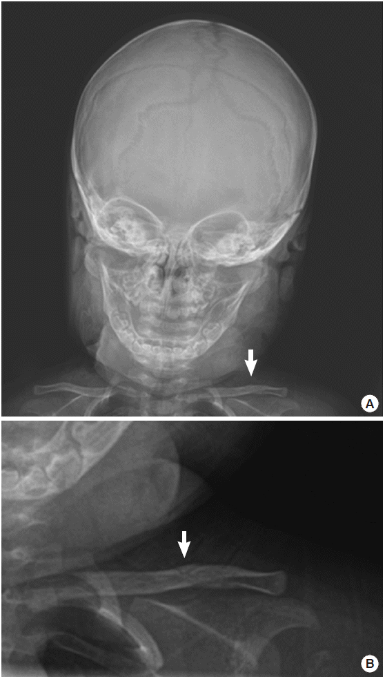
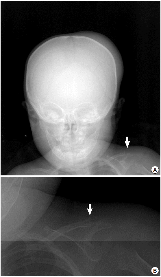
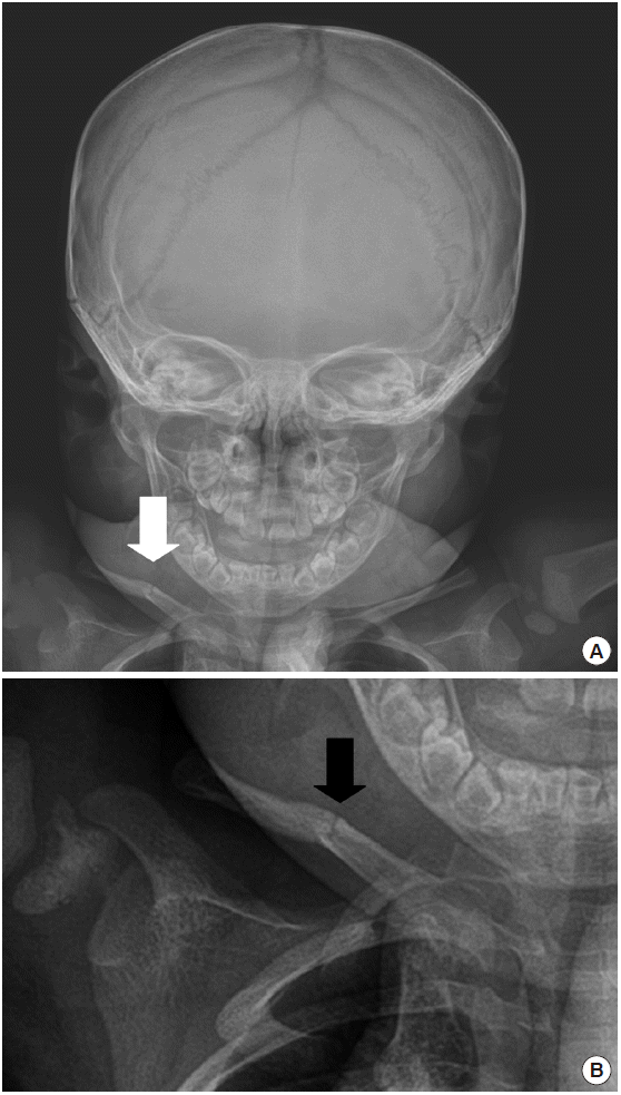
Tables 2 and 3 summarize characteristics of the patients with clavicle fractures. Of note, only 4 patients were accurately diag-nosed and only 2 patients had comments recorded by the pediat-ric radiologists (Table 3). We identified 3 non-displaced fractures, 1 cortical defect (patient G in Table 3, Fig. 2), 1 left acromiocla-vicular joint injury (patient H in Table 3, Fig. 3) and 1 greenstick fracture (patient J in Table 3, Fig. 4). The left acromioclavicular joint injury was regarded as a clavicle fracture (Fig. 3). Among 10 patients with clavicle fractures, no descriptions of complications were available for 6 patients and all but 1 patient were classified as missed. However, we did not perform follow-up examinations to determine complications or outcomes.
DISCUSSION
To our knowledge, this is the first study using a skull X-ray to identify clavicle fractures. Skull AP X-ray is a common radiologic modality for diagnosing skull fractures in the PED, whereas it only covers the skull and upper part of the cervical spine in adults, it includes a larger area in infants because of the high head-to-body ratio. Our hospital policy requires skull X-rays to be ordered for all patients with head injuries and these are usually taken as a 4-view series (anterior-posterior, both lateral, and TowneŌĆÖs views) although the ideal number of views is an ongoing subject of debate [3-5]. In our opinion, among 4 views, the clavicle is best recognized on a skull AP. On that view, both clavicles are usually clearly seen with minimal overlapped density caused by soft tissue or guardianŌĆÖs hands. In this study, 89.6% of patients had bilaterally visible clavicles (Fig. 1) although in 58.2% of patients, the clavicles were covered by the guardianŌĆÖs hands. The clavicles were discernible on most of these images, but it is possible that some non-displaced fractures were hidden.
Approximately 10% to 15% of all fractures in children involve the clavicle, but in one study infants accounted for only 1% to 2% of 537 cases of clavicle fractures [1,2]. In our study, 10 patients (1.4%) with clavicle fracture were identified, and 6 fractures (0.8%) were missed at the time of presentation. This suggests that diagnosing clavicle fractures on skull X-rays is not easy. Even when using a clavicle X-ray, non-displaced fractures can hinder a correct diagnosis. Jones et al. [6] reported that interobserver agreement in the classification of clavicular mid-shaft fractures was good for comminution (╬║ = 0.80, P < 0.001), but only moderate for displacement (╬║ = 0.76, P < 0.001) and operative treatment (╬║ = 0.64, P < 0.001). Missed fractures on clavicle X-rays have been described in a few previous case reports [7,8]. Of 3 patients with non-displaced fractures in our study (Figs. 2ŌĆō4), none had available comments written by pediatric radiologists, and 2 of the fractures were missed at initial presentation. Injuries to the lateral end of the clavicle in children are probably physeal injuries rather than dislocations because the physis fuses at 25 years and is weaker than the acromioclavicular ligament [9]. Accordingly, we regarded our case of left acromioclavicular joint injury as a fracture (Fig. 3).
Birth injuries and child abuse must be considered in infant cases. The clavicle is the most commonly injured bone during birth, and Kaplan et al. [10] reported that clavicle fractures develop in 1.65% of all vaginal deliveries. In our study, 1 birth injury occurred without a clavicle fracture. Karmazyn et al. [11] reported that clavicle fractures and skull fractures account for 2% and 7% of child abuserelated fractures, respectively. Given the high prevalence of skull fractures, skull APs are taken more frequently than clavicle X-rays. Van Laarhoven et al. [12] reported that a skull fracture is the most commonly associated injury in severely injured patients with clavicle fractures. Thus, physicians should maintain a high index of suspicion for clavicle fractures when they read a skull AP because of the frequency with which skull APs are performed and the association between skull and clavicle fractures. In our study, only 2 patients had both skull and clavicle fractures, and for 1 of them, the mechanism of injury was unknown (patient H in Table 3). Because the patient in question had a fracture of the lateral end of the clavicle, which is an injury with a high-specificity for child abuse [13], it is possible that child abuse was committed.
In this study, falls were the most common injury mechanism (86.0%, Table 1) and this was also true among the patients with clavicle fractures (8 of 10 patients in Table 3). Soto et al. [14] reported falls as the most common injury mechanism (50%) among 189 patients aged 2 years or younger. Because of the strict age range (1 year or younger) applied in our present analyses, this is the first study to report the incidence of clavicle fractures among infants with fall-related injuries.
Few complications are associated with clavicle fractures in children because of the remodeling potential of the clavicle [9]. Calder et al. [15] suggested that patients with uncomplicated clavicle fractures do not need routine follow-up, and Strauss et al. [1] reported that increasing age and complete displacement are predictive of complications. In this study, all patients were younger than 1 year and only 1 patient had a complete displacement (patient A in Table 3), thus, it is unlikely that they would develop complications. We did not perform further investigations because they would have been beyond the scope of the study. Of 6 patients with missed fractures in this study, descriptions regarding their complications were unavailable in all but 1 patient.
We offer the following suggestions to ensure that clavicle fractures are not missed. First, all skull X-rays of infants should be examined with the intent of examining the clavicle. Second, if the presence of a fracture is uncertain, the injury mechanism, tenderness or palpable lumps on the clavicle, and pseudoparalysis should be reevaluated. Third, given the high frequency of skull APs that include the clavicle (89.6%) and radiation hazard of additional X-rays, revision of the skull X-ray protocol to include assessment of the clavicle could be implemented. Fourth, ultrasound as a complementary modality could be used, especially when as-sessing non-displaced fractures. A previous prospective study, using ultrasound to diagnose clavicle fractures in children demonstrated 95% sensitivity and 96% specificity [16]. In another study in which most participants were novice ultrasonographers, 89.7% sensitivity and 89.5% specificity were reported [17]. Therefore, we favor using ultrasound to diagnose suspected fractures, however, additional specialized studies are required.
Our study has some limitations. First, this was a single-center retrospective study and we could not always determine if an accurate diagnosis had been made using a skull AP. However, we did evaluate all skull AP findings and diagnosed only the lesions that were recognizable as clavicle fractures using this modality. Second, the plain X-ray was the reference standard in our study though it has only moderate interobserver agreement according to investigators [6]. Although interobserver agreement is not strong for fractures without comminution, no interobserver disagreement was found in our study. This might be due to the small number of patients. Third, retrospective diagnosis of non-displaced clavicle fractures (e.g., greenstick fracture in Fig. 4) might be inaccurate, and the lack of reported clinical manifestations in the electronic medical records (e.g., tenderness, shoulder deformity) could have contributed to the inaccuracy. The lack of clinical data may have occurred because charting in head injury cases tends to be head injury-oriented. In addition, during the study period, ultrasound was not yet used as a standard protocol at our hospital. However, in the case of uncertain diagnoses, we made the final determinations after double-checking the records and discussing the cases. Fourth, we did not evaluate associated injuries, complications, or medico-legal aspects. However the specific purpose of this study was to investigate how often the clavicle and its fractures were recognizable on skull AP, and we considered the secondary issues less relevant. Finally, we tried, but failed to determine the factors associated with missed clavicle fractures because the number of missed cases was relatively small (6 of 734 cases, 0.8%).
We found that in 89.6% of the patients, skull APs identified bilaterally visible clavicles, and that 10 of these patients (1.4%) had clavicle fractures. In 6 of these patients (0.8%), the clavicle fractures were initially missed. Unfortunately, we could not determine the factors associated with a missed diagnosis. Clavicle fractures are easy to miss on skull X-rays, even when specifically looking for them, especially when unsuspected.


