INTRODUCTION
Water beads are spherical, gelatinous toys measuring 2 to 3 mm that expand on contact with water, particularly in alkaline pH [1]. Young children may be attracted to swallow the toys by their candy-like appearance and bright color [1,2]. The beads are radiolucent and difficult to detect on plain radiographs. In addition, young children typically cannot communicate well about the swallowing events. Thus, ingestion of water beads is prone to delayed diagnosis and consequent intestinal obstruction. Recently, a case was reported on the use of point-of-care ultrasound (POCUS) in an infant with esophageal obstruction caused by food [3]. This case shows the advantages of the imaging modality, including rapid applicability, repeatability, and avoidance of radiation and sedation. We aimed to describe the use of POCUS for early detection of multiple ingested water beads in an infant.
CASE REPORT
A previously healthy 12-month-old girl presented to the emergency department with non-bilious vomiting that had started nine hours before the visit. She was presumed to have gastroenteritis and was prescribed oral rehydration solution at a pediatrician’s office. Subsequently, she vomited six more times, and in two episodes the vomitus contained spherical, green, gelatinous toys that were about 3 mm in size. No choking or swallowing of the toys was witnessed. However, the girl’s mother believed that they were water beads and that some likely remained in the girl’s body given the disappearance of several beads at home.
The initial vital signs at the emergency department included blood pressure 90/70 mmHg, heart rate 140 beats/min, respiratory rate 32 breaths/min, and temperature 37.0°C. Her weight and height were 11.0 kg (90th to 95th percentile) and 75.3 cm (50th to 75th percentile), respectively. She had a stable and mildly anxious appearance without respiratory distress or dehydration. The abdomen was soft, non-distended, and non-tender without a palpable mass.
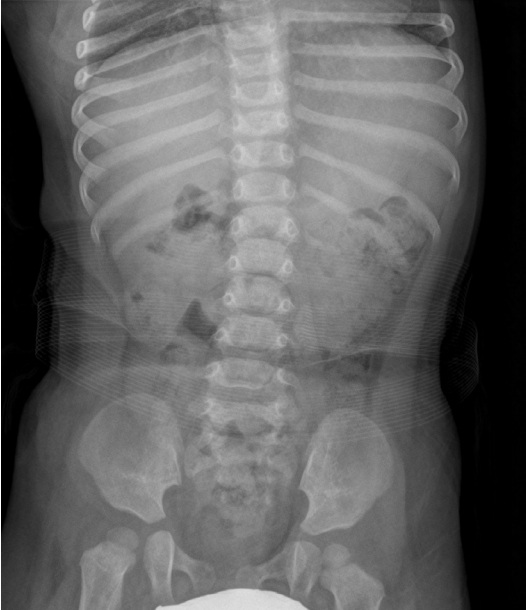
No intestinal obstruction or radiopaque foreign bodies were found on a plain abdominal radiograph (Fig. 1). However, considering the mother’s concern for residual foreign bodies, abdominal POCUS was performed using an HM70A ultrasound system (Samsung Medison, Seongnam, South Korea) to detect water beads. A 7-MHz curvilinear transducer was placed midline on the epigastrium with the patient supine and the indicator on her right side.
POCUS showed the stomach as a fluid-filled sac, indicating gastric distention due to distal intestinal obstruction or retention of rehydration solution. Moreover, seven well-demarcated, round hypoechoic items approximately 10 to 15 mm in diameter were observed in the stomach and a similar, 20 mm item in the first portion of the duodenum (Figs. 2, 3). No other foreign bodies, masses, dilated loops of bowel, or other signs of obstruction, such as intussusception, were observed on the examination. The POCUS findings indicated duodenal obstruction by ingested water beads. Of note, this examination was completed within several minutes without sedation, use of a linear transducer, or application of extra pressure on the abdomen.
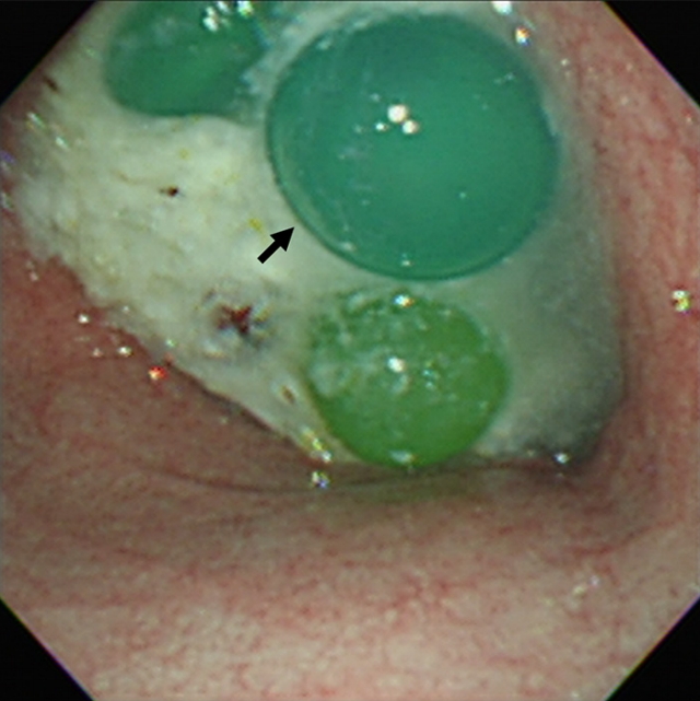
Subsequently, a pediatric gastroenterologist was able to retrieve the water beads endoscopically (Fig. 4). A total of 11 water beads were removed from the stomach using a retrieval net. The bead in the duodenum was first broken down by endoscopic crushing. The patient’s recovery was uneventful, and she was discharged on day 3.
DISCUSSION
Ingested water beads can be readily observed on POCUS as welldemarcated, round hypoechoic materials. Thus, they are visually ideal for detection by novice POCUS users who might otherwise hesitate to use the application in emergency settings, particularly in combination with a lack of fasting or cooperation in young children. In this case, the early detection was further expedited by the use of two acoustic windows, including the left lobe of the liver and the fluid-filled stomach with scanty gas.
The impact of water- and pH-sensitive expansion of water beads is obvious with POCUS [1]. Notably, separate studies have shown the diameter of the toys can increase from 2.0 to 9.5 mm and from 7.5 to 40.0 mm when immersed in water for 12 hours [2,4]. Swollen water beads may therefore obstruct the small bowel, which has a diameter of 25 to 30 mm. The bead size also increases with increasing pH [1,4]. This chemical change is exemplified by the larger size of the duodenal bead compared to the gastric ones in this case. The larger bead may be attributed to a longer contact time with water and the higher pH in the duodenum.
In this case, the use of POCUS expedited endoscopic intervention. Other reported cases of water bead ingestion have noted median delays in diagnosis of 3 days (range, 15 hours to 1 month) and the obstructions were more distal than in this case (Table 1) [1,2,5-8]. Of note, the diagnosis in this case was made before the toys passed the second part of the duodenum, which is the typical boundary of endoscopy. An ultrasound diagnosis of water bead ingestion has been reported in three cases. However, the diagnoses were made 2 days to 1 month after symptom onset, and all patients underwent surgical removal (Table 1) [1,2,5-8]. Despite the larger number of ingested water beads in our case, the early detection with POCUS prevented surgery, which is the usual procedure in such cases [2,5-8].
A couple of pitfalls were noted in using POCUS to detect ingested water beads. First, POCUS may underestimate the number of water beads when compared to endoscopic evaluation. These attempts should not delay endoscopic removal of foreign bodies, even though multi-directional or repetitive scans could compensate for two-dimensional sonography limitations. Second, examining the small bowel should be done carefully by discriminating the double-wall sign of water beads from enteric duplication cysts, both of which can often be confusing at the level of the small bowel.
Ingested water beads can be readily detected on POCUS given the physical and chemical features of the toys. In such cases, the use of POCUS could expedite endoscopic intervention and avoid surgical removal of foreign bodies.