| Home | E-Submission | Sitemap | Contact Us |  
Search
Clin Exp Emerg Med Search

CLOSE

Clin Exp Emerg Med > Volume 8(3); 2021 > Article
Lee, Lee, and Kim: Strangulated appendiceal intussusception caused by isolated endometriosis
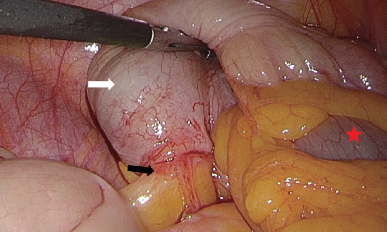
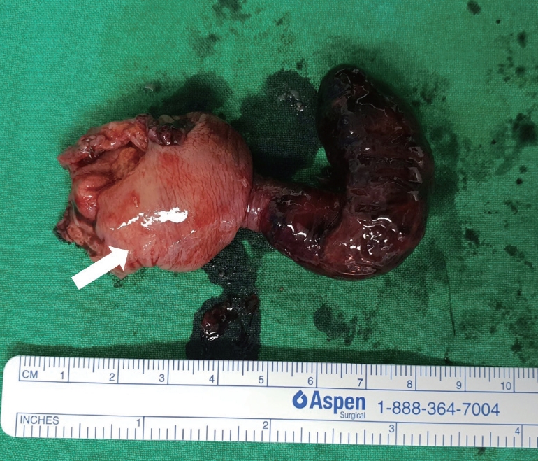
A 33-year-old female was admitted to our emergency department for right lower quadrant abdominal pain, which was not associated with her menstrual cycle, and nausea, which had persisted for 3 days. She denied any prior history of abdominal pain, dysmenorrhea, menorrhagia, or dyspareunia. The patient had no family history of endometriosis. Abdominal computed tomography revealed appendiceal intussusception (AI) and invagination of the periappendiceal fat into the cecum (Fig. 1). She underwent laparoscopic partial cecectomy with the presumed diagnosis of AI (Fig. 2). The ovaries and fallopian tubes were normal, with no endometrial spot. The inverted appendix was completely necrotic (Fig. 3). Final surgical pathology was positive for gangrenous intussusception of the appendix, with endometrial deposits (Fig. 4).
Adult colonic intussusception is caused by either malignant tumors (64%–87% cases) or benign tumors (29%–33%). The most common causes are adenocarcinomas and lymphomas among malignant tumors and lipomas, leiomyomas, adenomatous polyps, and endometriosis among benign tumors [1]. AI is extremely rare, with reported rates of 0.01% in patients who undergo appendectomy [2]. AI secondary to endometriosis is also extremely rare, with less than 30 cases reported. However, those cases involved endometriosis on other organs and the appendix. Only three cases involving isolated appendiceal endometriosis are reported in the literatures [3]. Patients with AI can range from being asymptomatic to presenting symptoms mimicking acute appendicitis [4]. Preoperative diagnosis is difficult owing to its rarity. Although endometriosis is reported to be the lead point of AI, there are limited descriptions on imaging findings. Computed tomography findings include an enlarged appendix with an inverted appendiceal tip or a tumor protruding into the cecum [5]. This case highlights that isolated endometriosis without involvement of reproductive organs can present as a strangulated AI. AI secondary to endometriosis should be considered in the differential diagnosis when female patients of childbearing age present with clinical symptoms of acute appendicitis and radiologic findings of intussusception, even before obtaining a pathological result.

NOTES

No potential conflict of interest relevant to this article was reported.

ACKNOWLEDGMENTS

This paper was supported by Wonkwang University in 2020.

REFERENCES

1. Lee SY, Park WC, Lee JK, Kang DB, Kim Y, Yun KJ. Laparoscopic treatment of adult sigmoidorectal intussusception caused by a mucinous adenocarcinoma of the sigmoid colon: a case report. J Korean Soc Coloproctol 2011; 27:44-9.
crossref pmid pmc
2. Collins DC. 71,000 Human appendix specimens. a final report, summarizing forty years’ study. Am J Proctol 1963; 14:265-81.
pmid
3. Manoharan B, Haider AS, Samira AM, Bharathan B, Parra-Blanco A. Isolated appendiceal endometriosis resulting in intussusception. Oxf Med Case Reports 2018; 10:349-52.
crossref
4. Salehzadeh A, Scala A, Simson JN. Appendiceal intussusception mistaken for a polyp at colonoscopy: case report and review of literature. Ann R Coll Surg Engl 2010; 92:W46-8.
crossref pmid
5. Hanly AM, Ryan EM, McNamara DA. Appendiceal intussusception and endometriosis mimicking colorectal cancer. Colorectal Dis 2011; 13:225-6.
crossref

Fig. 1.
The abdominal computed tomography. (A) Axial view and (B) coronal view. These images show a low-density tubular structure (5×1.6 cm in diameter) in the cecum near the appendiceal orifice, and focal invagination of the peri-appendiceal fat to the cecum (white arrow).
ceem-20-026f1.jpg
Fig. 2.
Surgical findings. During laparoscopic exploration, the appendix was not macroscopically visible in the right iliac fossa, but it was located in the cecum (white arrow), which showed no possibility for reduction (black arrow). Terminal ileum denoted by asterisk.
ceem-20-026f2.jpg
Fig. 3.
Surgical specimen. Appearance of the specimen resected by laparoscopic partial cecectomy. The inverted appendix (70×15 mm in size) is completely necrotic at the appendiceal orifice (inside the aspect of the cecum denoted by a white arrow).
ceem-20-026f3.jpg
Fig. 4.
High-power view of endometriosis within the appendix. Endometrial glands and stroma within the muscular layer, around the appendicular mucosa (hematoxylin and eosin, ×200).
ceem-20-026f4.jpg
TOOLS
PDF Links  PDF Links
PubReader  PubReader
ePub Link  ePub Link
XML Download  XML Download
Full text via DOI  Full text via DOI
Download Citation  Download Citation
  Print
Share:      
METRICS
0
Crossref
0
Scopus
2,588
View
65
Download
Editorial Office
The Korean Society of Emergency Medicine
101-3104, Brownstone Seoul, 464 Cheongpa-ro, Jung-gu, Seoul 04510, Korea
TEL: +82-31-709-0918   E-mail: office@ceemjournal.org
About |  Browse Articles |  Current Issue |  For Authors and Reviewers
Copyright © by The Korean Society of Emergency Medicine.                 Developed in M2PI