INTRODUCTION
Internal jugular vein catheterization (IJC) is useful in emergency departments (ED) for monitoring hemodynamic status and for infusion of fluid or nutrients. As compared to subclavian vein catheterization, IJC has fewer complications such as pneumothorax or hemothorax, and thus is more often recommended [1].
As first described in the 1980s, the use of bedside ultrasonography by emergency physicians in the ED has many advantages. For example, it can be performed 24 hours a day and is easily accessible. In addition, it can contribute to improved patient satisfaction and quality of emergency care, as well as a reduction in the length of ED stays [2].
In recent years, USG-guided IJC has been widely used. However, it may be difficult to perform in cases of small cross-sectional area (CSA) of the internal jugular vein (IJV) with USG-guidance (e.g., in hypovolemic patients). To overcome this limitation, methods such as the Valsalva maneuver, Trendelenburg position, and passive leg elevation have been applied to dilate the IJV [3-5]. The Valsalva maneuver is performed through moderately forceful attempted exhalation against a closed airway, usually done by closing one’s mouth while simultaneously expiring against a closed glottis. This elicits a cardiovascular response, increasing venous pressure and resulting in venous dilation.
Bellazzini et al. [6] reported that the Valsalva maneuver could dilate the IJV by 38%, or even more if combined with the Trendelenburg position. However, the Valsalva maneuver may be difficult to perform in unconscious patients or uncooperative patients such as children.
To our knowledge, this study provides the first description regarding digital proximal compression of the IJV. We aimed to investigate the effectiveness of digital proximal compression of the IJV on CSA-IJV alone and in combination with Valsalva maneuver.
METHOD
Study design
We conducted an experimental study to assess the effects of compressing the IJV on CSA-IJV. The study was approved by the hospital’s institutional review board, and informed consent was waived because the procedure is not invasive, not painful, and not time-consuming (AJIRB-MED-DEO-15-239).
Study population
We enrolled healthy volunteers for study participation. Employees of a tertiary hospital ED were recruited. We then excluded participants with a history of IJC or neck surgery.
Study protocol
Each patient lay down in the supine position, and the diameter of his or her neck was measured. The measurement point of the CSA-IJV was at the top point of the triangle created by the clavicle and the sternal and clavicular heads of the sternocleidomastoid muscle. By ultrasonography (L15W probe; Zonare Medical Systems, Mountain View, CA, USA), this was approximately at the level of the thyroid cartilage prominence.
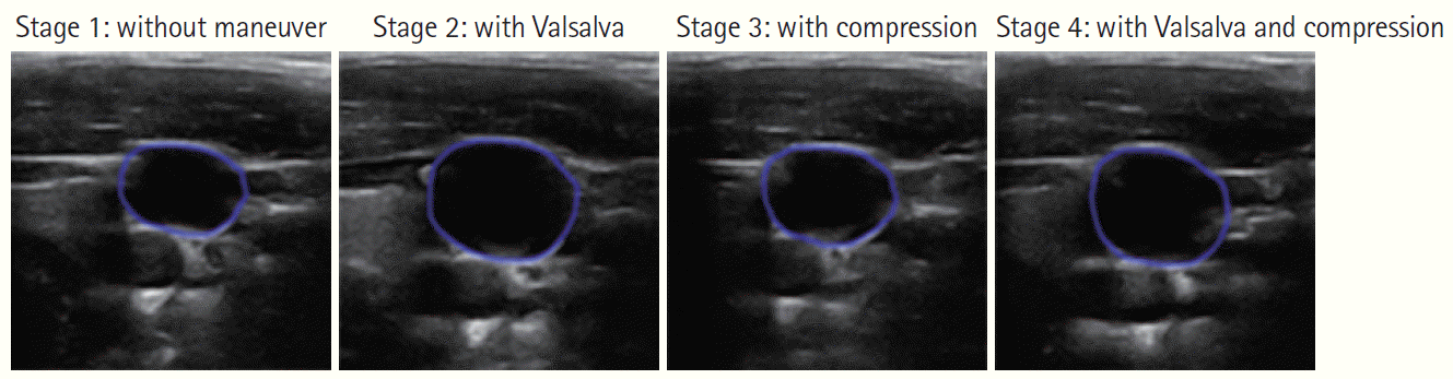
Three examiners independently measured the CSA-IJV. These examiners were all board-certified emergency physicians with academic experience with emergency ultrasonography. The average measurement on maximal diameter, our main outcome, was calculated by the ultrasonography machine (Fig. 1).
For each participant and examiner, the CSA-IJV was measured in four different stages. For the first stage, the CSA-IJV was measured with the participant at rest without any maneuver. For the second stage, the participant was instructed to perform the Valsalva maneuver by holding his or her breath and compressing the lower abdomen for 5 seconds, after which the CSA-IJV was measured. For the third stage, our main focus of the study, the participant was instructed to breathe normally while their proximal IJVV was compressed by the examiner’s index finger (Fig. 2). The compression point was identified as the lower border of the triangle that is delineated by the sternocleidomastoid and clavicle. The CSA-IJV was measured within 20 seconds of compression. The last stage combined techniques used in the second and third stages, such that digital proximal compression and the Valsalva maneuver were performed simultaneously. The CSA-IJV was measured within 20 seconds of compression. A five-minute resting period was allowed between each stage.
Statistical analysis
Variables were expressed as means and standard deviations. To test the normality, the Kolmogorov-Smirnov test was performed. Paired t-test was performed for comparison between groups. P-values <0.05 were considered significant. All analyses were performed using IBM SPSS ver. 22.0 (IBM Corp., Armonk, NY, USA) and R ver. 3.1.2 (R Foundation for Statistical Computing, Vienna, Austria; http://www.R-project.org/) software packages. The sample size was calculated from preliminary data using the ‘sample size’ library with R package. To detect a 0.2 cm2 differences with a power of 0.8 and a significance level of 0.05 from data with a standard deviation of 0.4 using a paired t-test, the required number of cases was calculated to be 39.
RESULTS
Baseline characteristics
A total of 41 volunteers were enrolled. Twenty-six (63.41%) were male, and the mean age was 28.15±2.85 years. Mean height was 170.74±8.66 cm and mean neck circumference was 35.28±3.87 cm. One volunteer had a history of neck surgery and was excluded from the study. Two participants had tonsillectomies prior to the study, but were included nonetheless. The general characteristics of participants are presented in Table 1.
Comparison of outcomes
The means and standard deviations of CSA-IJV following each maneuver are listed in Table 2.
The CSA-IJV with the Valsalva maneuver was significantly greater than that of the Stage 1 control (1.34±0.45 vs. 1.06±0.36 cm2, respectively, P<0.001). The CSA-IJV with digital proximal compression of the IJV was significantly different from that of the control (1.26±0.41 vs. 1.06±0.36 cm2, respectively, P<0.001). Moreover, the CSA-IJV under Valsalva maneuver was significantly larger than the CSA-IJV with proximal compression of the IJV (1.34±0.45 vs. 1.26±0.41 cm2, respectively, P=0.0112).
The CSA-IJV during simultaneous digital proximal compression and the Valsalva maneuver was significantly larger than that of digital compression alone (1.41±0.47 vs. 1.26±0.41 cm2, respectively, P<0.001). It was also larger than that of the Valsalva maneuver alone, but without achieving statistical significance (1.41±0.47 vs. 1.34±0.45 cm2, respectively, P=0.062).
DISCUSSION
To our knowledge, this is the first study to show the effectiveness of digital proximal compression of the IJV on the CSA-IJV.
For many years, the blind IJC method has been used in the ED. However, blind IJC might lead to serious complications, such as arterial puncture, nerve injury, or failure. As a result, ultrasound-guided IJC is currently the method of choice.
Many modifications have been suggested to improve the success of IJC. Wang et al. [7] reported that the angle of the neck or ultrasonography-guided method could affect the success rate of IJC. Armstrong et al. [8] showed that use of the Trendelenburg position up to 30 degrees enlarges the CSA-IJV and increases the success rate of IJC. Other methods, such as compression of the liver or placement of a pillow under the head, have been investigated. On the other hand, compression of the carotid artery or a pillow under the shoulder have been found to lower the success rate of IJC [9,10].
The Valsalva maneuver could be an ideal method to increase the CSA-IJV, and thereby facilitate IJC, but it requires the cooperation of patients. Therefore, it definitely could have limitations in pediatric or unconscious patients. The Trendelenburg method can be performed on uncooperative patients, but requires a tilting table, which may not be available in all EDs around the world.
Given these limitations, proximal compression of the IJV, which could be easily done in any ED, might be useful in performing IJC. In this study, even though the CSA from proximal compression of the IJV was slightly smaller than that following the Valsalva maneuver, it was much larger than the control CSA-IJV. The amount of dilation achieved could effectively aid proper insertion of an IJV line. Moreover, the combination of digital proximal compression and the Valsalva maneuver resulted in the largest CSA-IJV. This suggests that ED physicians could perform proximal compression alone in uncooperative patients to improve the odds of successful IJC. In cooperative patients, they could combine this technique with the Valsalva maneuver to help maximize the CSA-IJV.
We have a few limitations to discuss. First, in this study, we simply measured the CSA-IJV with each maneuver. Due to ethical considerations, we could not perform the IJC and compare the success rates of this invasive procedure. However, we can safely infer that a larger CSA-IJV would enhance the odds of success in IJC. Second, we could not standardize the digital pressure on the proximal IJV. In reality, it would be hard or nearly impossible for clinicians to exert the same pressure. Finally, our statistical significance could be strengthened by enlarging the study sample size.
In summary, digital proximal compression of the IJV dilated the IJV, and this could have a positive effect on the success of IJC. Further clinical trials are needed to validate this technique.