| Home | E-Submission | Sitemap | Contact Us |  
Search
Clin Exp Emerg Med Search

CLOSE

Clin Exp Emerg Med > Volume 10(2); 2023 > Article
In, Oh, and Min: Hyperacute presentation of fat embolism syndrome after multiple long bone fractures
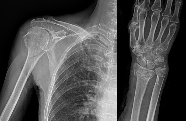
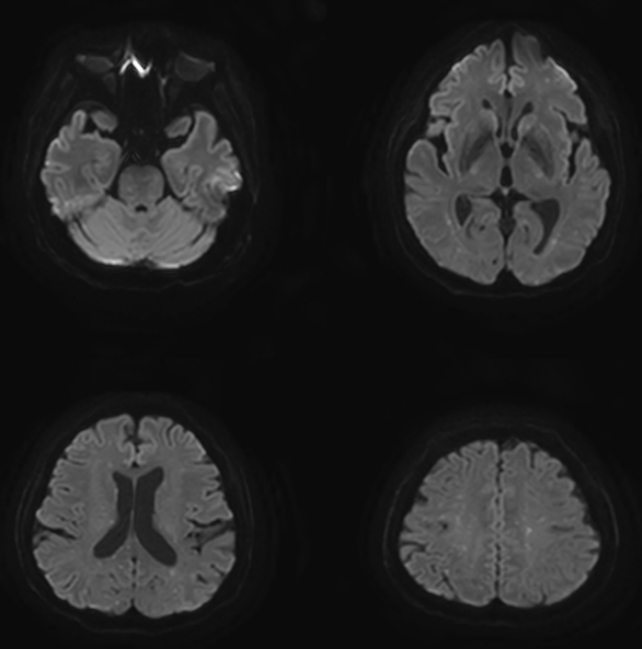
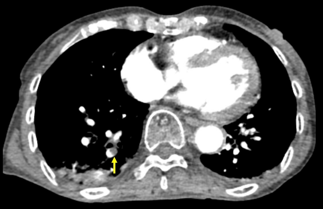
A 69-year-old woman presented to the emergency room with right arm pain after a fall. She was alert and had no other symptoms. She had no relevant medical history. The radiograph showed a right distal radius fracture and fracture of the surgical neck of the humerus (Fig. 1). After 1 hour, she had a sudden change in consciousness, with no response to pain stimulus. Brain computed tomography (CT) was performed immediately but showed no intracranial hemorrhage. Subsequent brain magnetic resonance imaging (MRI) showed multiple embolic infarcts (Fig. 2). In addition, chest CT showed pulmonary thromboembolism (Fig. 3). A second brain MRI performed 2 days after hospitalization showed multiple embolic infarcts with progression (Fig. 4). No evidence of an embolic source was found in the electrocardiogram and echocardiography. The patient remained in a state of stupor mentality throughout the 2-month inpatient treatment. Additionally, the electroencephalogram examination confirmed the presence of epilepsy. After undergoing conservative treatment for 2 months, the patient was transferred to a rehabilitation hospital.
Although histological confirmation was not performed, sudden neurologic deficits, tachypnea, and disseminated intravascular coagulation after long bone fractures strongly suggested fat embolism syndrome (FES), a rare and fatal complication that can occur after a long bone fracture [13]. FES usually occurs between 12 and 72 hours postfracture and rarely occurs before 12 hours [2]. This patient developed FES 90 minutes after injury, which is very rare. Neurological symptoms or signs of cerebral FES vary widely, ranging from headache, diffuse encephalopathy, aphasia, and seizures [4,5]. FES should be considered in the differential diagnosis of patients with sudden changes in mental status immediately after a long bone fracture.

NOTES

ETHICS STATEMENTS
Informed consent for publication of the research details and clinical images was obtained from the patient.
CONFLICT OF INTEREST
No potential conflict of interest relevant to this article was reported.
FUNDING
None.
AUTHOR CONTRIBUTIONS
Conceptualization: SKO; Data curation: YNI, SKO; Investigation: SKO; Methodology: JHM; Resources: YNI; Software: JHM; Supervision: SKO; Validation: SKO; Visualization: YNI, SKO; Writing–original draft: JHM, SKO; Writing–review & editing: all authors. All authors read and approved the final manuscript.

REFERENCES

1. Chang RN, Kim JH, Lee H, et al. Cerebral fat embolism after bilateral total knee replacement arthroplasty: a case report. Korean J Anesthesiol 2010; 59 Suppl(Suppl):S207-10.
crossref pmid pmc
2. Huang CK, Huang CY, Li CL, et al. Isolated and early-onset cerebral fat embolism syndrome in a multiply injured patient: a rare case. BMC Musculoskelet Disord 2019; 20:377.
crossref pmid pmc pdf
3. Jorens PG, Van Marck E, Snoeckx A, Parizel PM. Nonthrombotic pulmonary embolism. Eur Respir J 2009; 34:452-74.
crossref pmid
4. Habashi NM, Andrews PL, Scalea TM. Therapeutic aspects of fat embolism syndrome. Injury 2006; 37 Suppl 4:S68-73.
crossref pmid
5. Kellogg RG, Fontes RB, Lopes DK. Massive cerebral involvement in fat embolism syndrome and intracranial pressure management. J Neurosurg 2013; 119:1263-70.
crossref pmid

Fig. 1.
Initial x-ray showing multiple bone fractures, including right distal radius and the surgical neck of the humerus.
ceem-22-419f1.jpg
Fig. 2.
Magnetic resonance imaging 2 hours after injury demonstrating findings of tiny, multifocal, diffusion-restrictive lesions in both the cerebrum and cerebellum.
ceem-22-419f2.jpg
Fig. 3.
Pulmonary artery thromboembolism in sub-subsegmental pulmonary arteries in the right lower lung (arrow). The embolus site was 12 Hounsfield units.
ceem-22-419f3.jpg
Fig. 4.
Magnetic resonance imaging 2 days after injury shows an increased number of tiny, multifocal, diffusion-restrictive lesions in the cerebrum, midbrain, pons, and cerebellum.
ceem-22-419f4.jpg
TOOLS
PDF Links  PDF Links
PubReader  PubReader
ePub Link  ePub Link
XML Download  XML Download
Full text via DOI  Full text via DOI
Download Citation  Download Citation
  Print
Share:      
METRICS
1
Web of Science
1
Crossref
0
Scopus
1,105
View
119
Download
Editorial Office
The Korean Society of Emergency Medicine
101-3104, Brownstone Seoul, 464 Cheongpa-ro, Jung-gu, Seoul 04510, Korea
TEL: +82-31-709-0918   E-mail: office@ceemjournal.org
About |  Browse Articles |  Current Issue |  For Authors and Reviewers
Copyright © by The Korean Society of Emergency Medicine.                 Developed in M2PI