| Home | E-Submission | Sitemap | Contact Us |  
Search
Clin Exp Emerg Med Search

CLOSE

Clin Exp Emerg Med > Volume 11(1); 2024 > Article
Gottlieb, O’Brien, Ferrigno, and Sundaram: Point-of-care ultrasound for airway management in the emergency and critical care setting

Abstract

Airway management is a common procedure within emergency and critical care medicine. Traditional techniques for predicting and managing a difficult airway each have important limitations. As the field has evolved, point-of-care ultrasound has been increasingly utilized for this application. Several measures can be used to sonographically predict a difficult airway, including skin to epiglottis, hyomental distance, and tongue thickness. Ultrasound can also be used to confirm endotracheal tube intubation and assess endotracheal tube depth. Ultrasound is superior to the landmark-based approach for locating the cricothyroid membrane, particularly in patients with difficult anatomy. Finally, we provide an algorithm for using ultrasound to manage the crashing patient on mechanical ventilation. After reading this article, readers will have an enhanced understanding of the role of ultrasound in airway management.

INTRODUCTION

Rapid airway management is a critical component of care in the acutely ill and decompensating patient. National data suggest that there are nearly 400,000 intubations performed in the emergency department (ED) each year [1]. These intubations are at increased risk of complications due to their emergent nature [2]. One large multicenter trial of intubations among the critically ill [3] reported severe complications (i.e., hypoxia, hypotension, or cardiac arrest) in 18% of patients. Another ED-based study [4] reported a 12% adverse event rate, including events such as esophageal or mainstem intubation, hypoxia, and cardiac arrest. These event rates are likely even higher among patients with difficult airways or those in cardiac arrest [2].
The use of point-of-care ultrasound (POCUS) for emergency procedures to supplement or transform conventional techniques is increasing rapidly [59]. Therefore, it is not surprising that this has also extended to airway management [10,11]. Ultrasound can be used to assess for a difficult airway prior to intubation, allowing time for appropriate preparation and secondary plans for airway management. Additionally, POCUS can be used to confirm proper placement and depth of the tube. After intubation, ultrasound can be utilized to assess and identify problems preventing adequate ventilation. In the event of a known difficult intubation, POCUS can be leveraged to accurately identify the cricothyroid membrane even when external anatomy is challenging for palpation.
In this article, we will outline the application of ultrasound for airway assessment, confirmation, and management as well as provide guidelines for troubleshooting a decompensating ventilated patient.

ASSESSMENT OF THE DIFFICULT AIRWAY

Up to 90% of difficult airways are unanticipated [12]. The management of the difficult airway presents a significant challenge in clinical practice, and as such, various tools and techniques have been developed to help make accurate assessments prior to intubation. Traditionally, this has been done using risk factors and physical examination maneuvers. Numerous assessment tools have been reported with variable diagnostic accuracy [13]. The upper lip bite test is often considered to have the best diagnostic accuracy, though one study found that the sensitivity was only 67% [1416]. Another commonly used tool, the modified Mallampati classification, had a sensitivity of 53% and a specificity of 89% [16].
Having the ability to assess the airway prior to intubation is a valuable skill and one that should be done quickly and accurately. POCUS is a helpful adjunct in airway assessments and can be done rapidly and noninvasively. There are several key measurements that can be used to predict difficult airways.
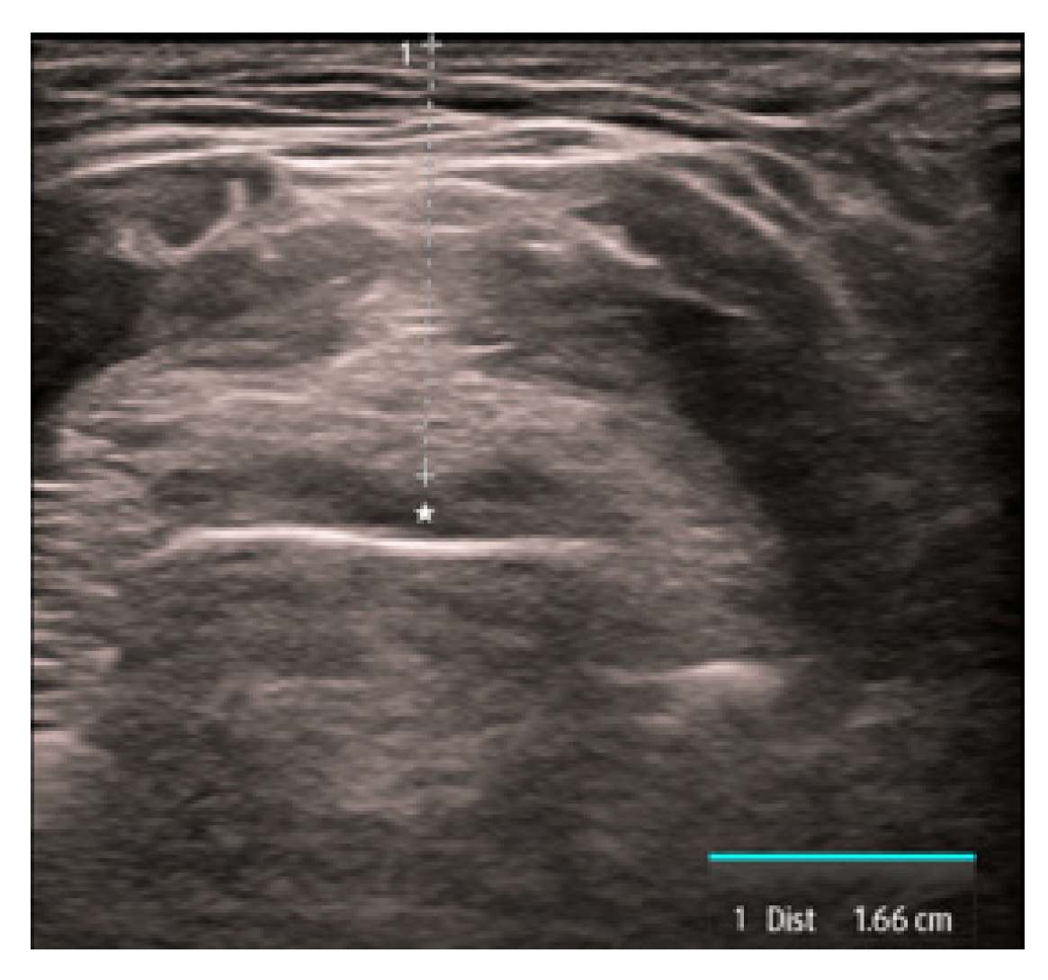
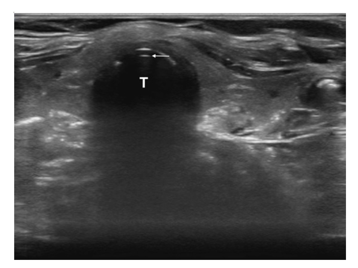
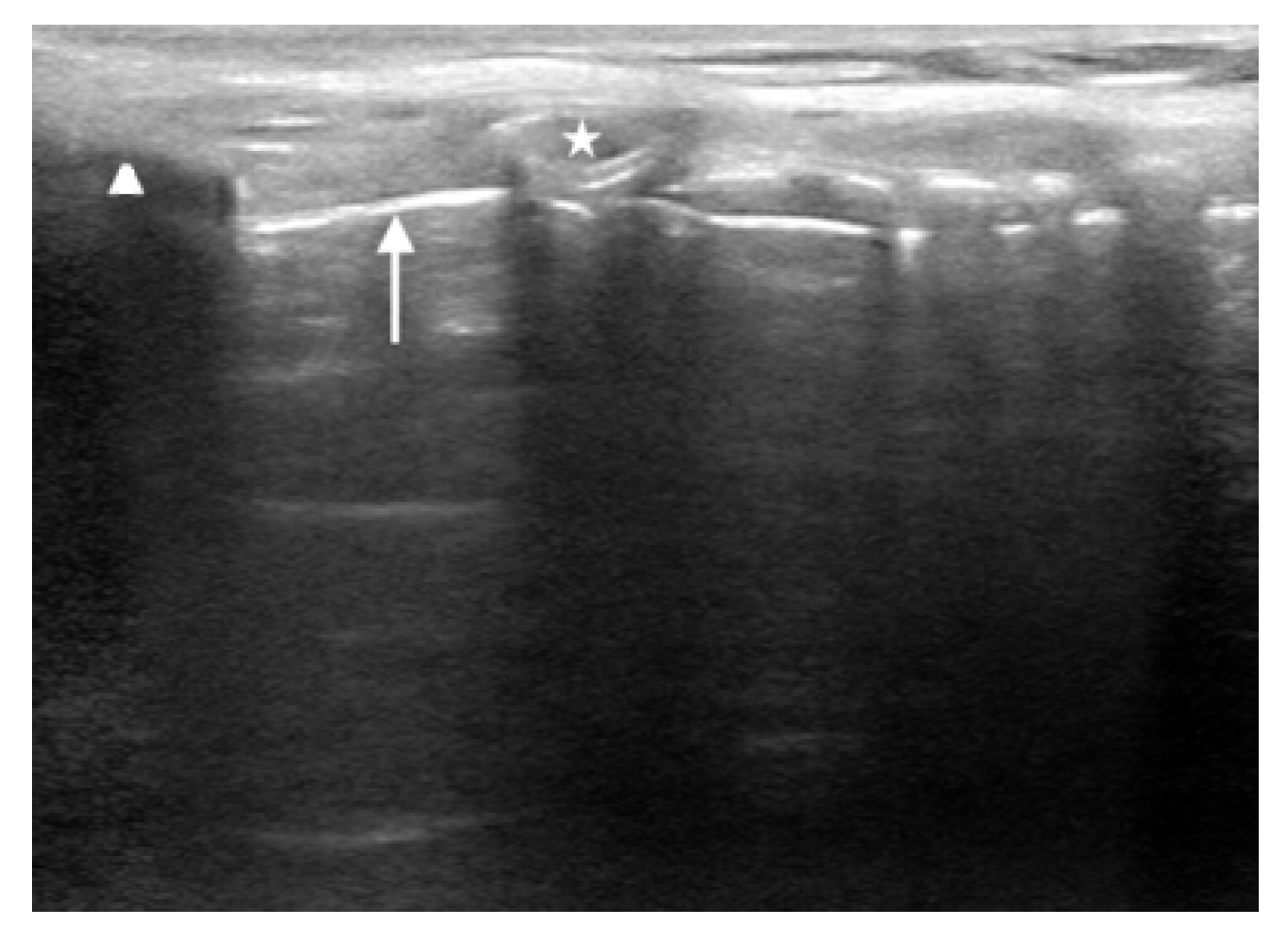
The first is the distance from the skin to the epiglottis (DSE) [1722]. This measurement is taken at the level of the thyrohyoid membrane. The linear transducer is placed transversely in the midline just superior to the thyroid cartilage. The epiglottis is seen just deep to the thyrohyoid membrane and preepiglottic space and will appear as a hypoechoic linear structure above the air-mucosa interface (Fig. 1). The distance is then taken from the superficial skin edge to the anterior edge of the epiglottis. In a recent systematic review and meta-analysis [22], a DSE of ≥2.54cm had a sensitivity of 82% and a specificity of 91% for predicting a difficult airway.
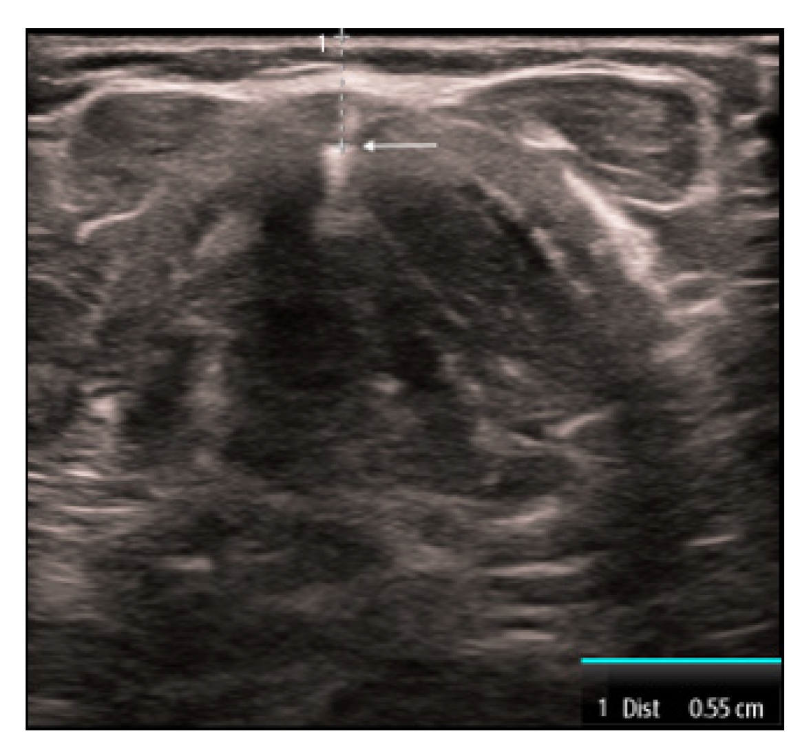
The second measurement is the distance from the skin to vocal cord (DSVC). This measurement is obtained by placing the linear transducer transversely in the midline over the thyroid cartilage. The strap muscles are seen anteriorly to the thyroid cartilage and the vocal cords are visualized just deep to the thyroid cartilage. The measurement is taken from the skin to the anterior commissure where the vocal cords join together (Fig. 2). A recent systematic review and meta-analysis [22] found that the DSVC had an overall sensitivity of 75% and a specificity of 72%. However, the literature is controversial with variable thresholds used and some even reporting an inverse association [23,24]. Therefore, the utility of this measure remains limited.
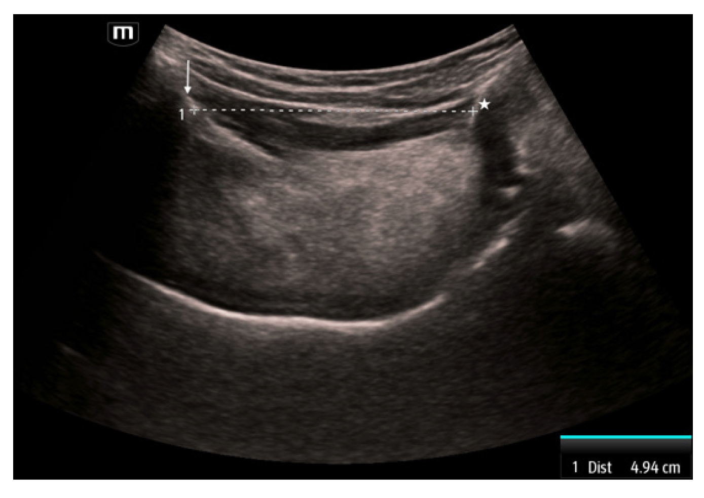
The third measurement is the hyomental distance (HMD), which is measured between the mentum of the mandible and the hyoid bone [21]. This measurement is obtained by placing the curvilinear transducer sagittally in the midline with the superior portion abutting the mandible. The mentum is seen as a hyperechoic structure with shadowing behind it on the superior portion of the image. The hyoid is a hyperechoic structure with shadowing behind it in the inferior portion. The measurement is taken between these two structures (Fig. 3). This distance has been reported as a single HMD, as a ratio of the HMD measured with the head in a ramped position compared to the head in neutral position (HMDR1), and the ratio of the HMD measured with the head in maximal extension compared to the head in neutral position (HMDR2) [21]. Patients with a shorter HMD in neutral position (<4.0 cm) were more likely to have a difficult airway [25]. Using a cutoff of 5.29 cm, HMD in the neutral position was 96.7% sensitive and 71.6% specific [26]. HMDR1 has less diagnostic utility with a 75% sensitivity and 76.2% specificity when using a threshold of 1.12 [27]. In contrast, HMDR2 has been reported to be 100% sensitive and 90.5% specific when using a threshold of 1.23 [27].
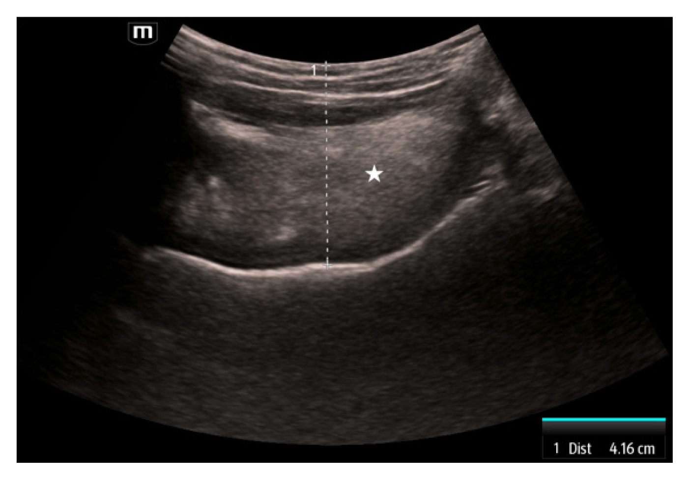
The fourth measurement is the tongue thickness. The tongue is visualized using a linear transducer placed transversely underneath the chin. The tongue is measured in the midline at the widest diameter in a superficial to deep direction beginning at the skin (Fig. 4). Tongue thickness has been demonstrated to be greater in difficult versus easy laryngoscopy (6.1–6.2 cm vs. 5.3–5.8 cm) [28,29]. When using a threshold of 6.1 cm, tongue thickness is 71% to 75% sensitive and 72% specific [28,29].
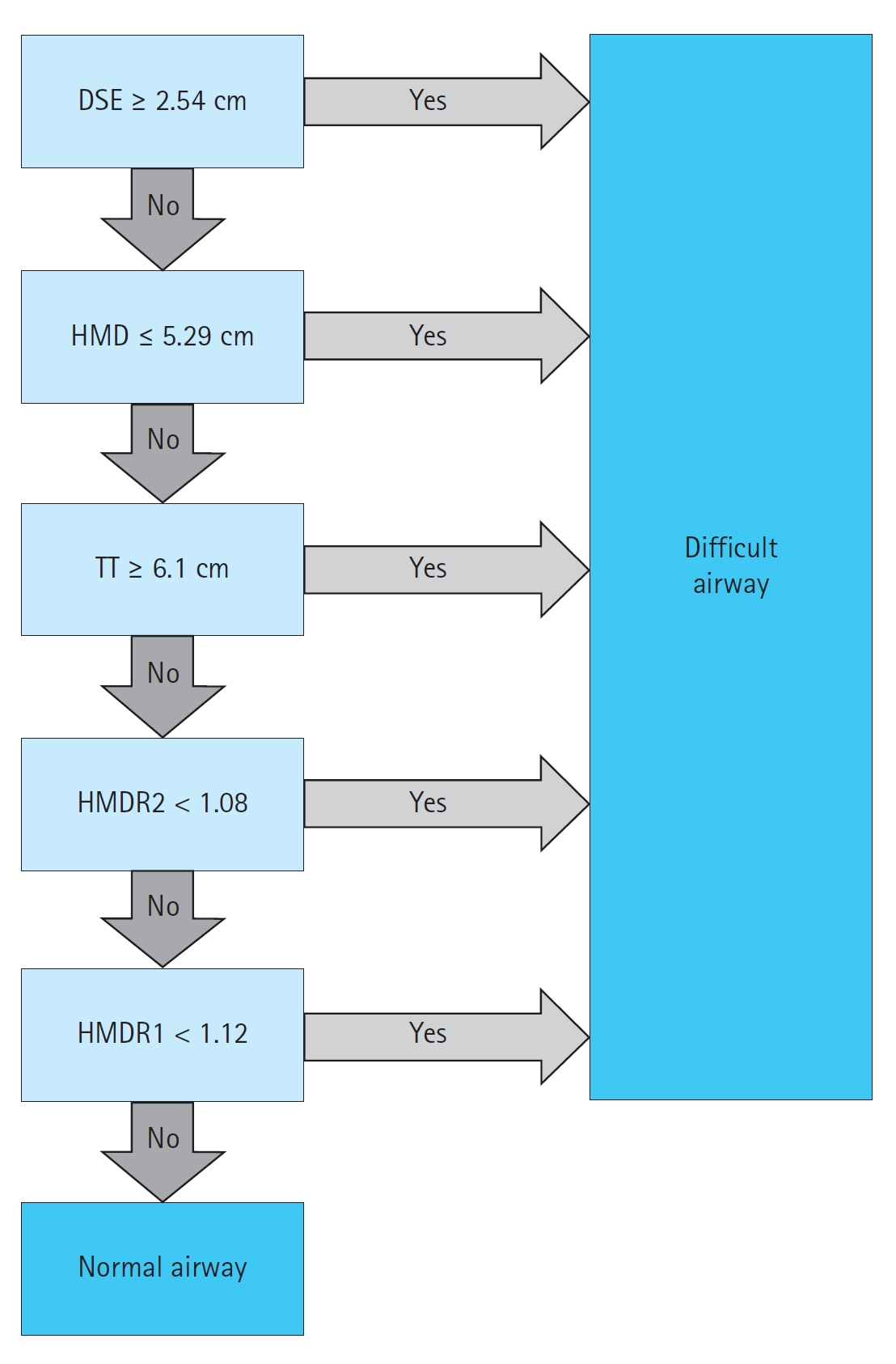
Lin et al. [21] proposed a protocol for this assessment, the Difficult Airway Evaluation with Sonography (DARES). The DARES protocol uses DSE, tongue thickness, HMD, HMDR1, and HMDR2 to predict a difficult airway (Fig. 5). An airway is considered difficult if any of the findings are positive. While informed by existing literature, this algorithm has not been externally validated.

INTUBATION CONFIRMATION

After the intubation is performed, it is critical to confirm that the endotracheal tube (ETT) was correctly placed in the trachea. Data suggest that the first-pass success rate for endotracheal intubation is 84% in the ED and 78% in the prehospital environment [30,31]. Confirmation of ETT location typically involves direct visualization of the ETT passing through the vocal cords followed by one or more confirmatory techniques. Auscultation of bilateral breath sounds, visualizing misting of the ETT, or using an esophageal detector device have limited diagnostic accuracy for confirming ETT placement [11,3237].
While waveform capnography is more accurate than the aforementioned approaches, it can be limited by false positives in the case of hypopharyngeal placement or recent ingestion of a carbonated beverage, as well as false negatives when expired CO2 levels are low (e.g., flash pulmonary edema, massive pulmonary embolism, or cardiac arrest) [11,20,38,39]. Importantly, data suggest that the accuracy of end-tidal capnography in cardiac arrest may be as low as 64% [3335]. Capnography also requires several positive pressure ventilatory attempts, which can increase gastric distension and increase the risk for aspiration.
In contrast, POCUS can allow rapid confirmation during or after the intubation attempt with a relatively short training period [10,40]. Among adults, POCUS is 99% sensitive and 97% specific [10,41]; whereas, POCUS is 92% to 100% sensitive and 100% specific in pediatric patients [42]. Studies have reported consistent accuracy regardless of the ETT size or type of transducer (e.g., linear, curvilinear) used [43,44].
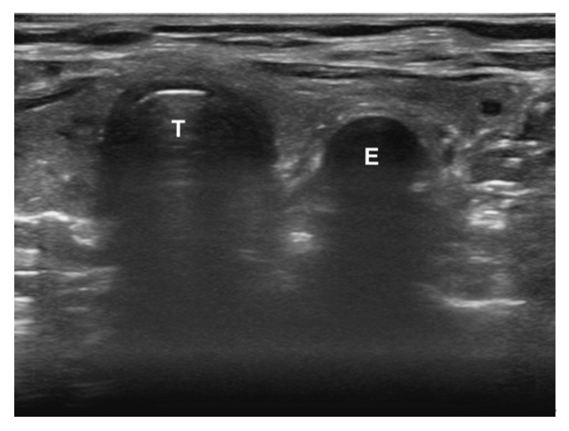
To confirm the ETT location, begin by placing the ultrasound transducer across the trachea in a transverse orientation. The ideal location is just superior to the suprasternal notch, as this has been shown to have superior visualization and diagnostic accuracy compared with other locations [45,46]. Confirmation can be performed in real-time (i.e., dynamic) or after the intubation (i.e., static). With the dynamic technique, the sonographer is assessing for rapid flutter-like movement as the ETT passes through the vocal cords (often referred to as the “snowstorm sign”) [20]. The static technique assesses for the appearance of either a thin hyperechoic membrane just posterior to the trachea (i.e., tracheal intubation) (Fig. 6) or a second air-mucosa interface referred to as the “double tract” sign (i.e., esophageal intubation) (Fig. 7) [20].
The literature has not demonstrated a significant difference in the accuracy between the static and dynamic techniques, so either are acceptable [10,47]. However, the static technique offers several unique benefits, including only requiring a single clinician (i.e., the intubator can also perform the POCUS immediately after the intubation is performed) and not placing the transducer on the neck during the intubation, which could make the intubation more challenging [20]. The major disadvantage is that it can be more difficult to locate the ETT behind the trachea due to the air artifact. In fact, data have shown reduced confidence and longer time to identification when the ETT is endotracheal versus esophageal [48]. To address this, some authors recommend twisting the ETT between one’s fingers to induce a false motion artifact [49,50]. While there was no significant difference in overall accuracy, ETT twisting has been shown to improve confidence and time to ETT identification [49]. Others have proposed infusing the ETT cuff with saline instead of air to better visualize the cuff [5153] .
In addition to direct visualization with transtracheal ultrasound, indirect signs such as lung sliding or diaphragmatic excursion can also be used to confirm ETT location. Bilateral lung sliding has been reported to be 92% to 100% sensitive and 56% to 100% specific [5459]. Similarly, diaphragmatic motion has 91% to 100% sensitivity and 50% to 100% specificity [6062]. When combined with transtracheal ultrasound, several studies [56,57] have found that the addition of lung sliding improves the accuracy compared with transtracheal ultrasound in isolation.

ASSESSMENT OF ETT DEPTH AND UNILATERAL LUNG INTUBATION

After confirming that the ETT is within the trachea, the next step is to assess the depth. If the ETT is placed too shallow, it can cause damage to the vocal cords or become dislodged [6365]. If the ETT is placed too deep it risks barotrauma in the ventilated lung along with atelectasis and hypoxia in the unventilated lung [65,66]. Data suggest that incorrect ETT depth can occur in up to 15% of adults and 18% of children [67,68].
Clinical markers like chest rise and breath sounds have limited accuracy for mainstem intubation and cannot provide any insight into the ETT depth [6971]. While chest radiography is considered the gold standard for ETT depth, it can take a significant amount of time, exposes the patient to radiation, and may delay other aspects of care in a critically ill patient [72].
In contrast, POCUS can provide rapid assessment of ETT positioning and assess for signs of mainstem intubation. In adults, transtracheal ultrasound had 84.8% accuracy for determining ETT depth and took on average 19 seconds to perform [73]. A recent systematic review and meta-analysis [74] found that ultrasound had 86.7% accuracy for detecting mainstem intubations, with a sensitivity of 93.0% and a specificity of 75.0%. In pediatric patients, one study [75] used a three-point technique (transtracheal ultrasound plus bilateral lung sliding) and reported 85.7% sensitivity and 98.3% specificity for mainstem intubation. Another study [76] used a saline-filled ETT cuff and was able to correctly identify ETT depth in 95% of cases.
To assess the depth of the ETT, start by placing the ultrasound transducer just superior to the suprasternal notch in the sagittal plane. Identify the thyroid cartilage, cricoid cartilage, and tracheal rings as hyperechoic, rounded structures in the near field with gray shadows. The ETT cuff will appear as a second, hyperechoic line directly posterior to these structures (Fig. 8). Saline can be instilled to improve visualization of the ETT cuff. If the cephalad border of the ETT cuff is seen at or above, the first tracheal ring it is considered too high; between the second and eighth tracheal rings is considered adequate; and below the eighth ring or not visualized is considered too deep [73]. One additional benefit of POCUS is real-time assessment. If the ETT is visualized either above or below the ideal location, it can be advanced or retracted using real-time guidance.
If there is any concern for mainstem intubation or the ETT cuff cannot be adequately visualized, the transducer should be placed longitudinally on the anterior chest at the midclavicular line to assess for lung sliding bilaterally [5459,75]. Unilateral lung sliding may suggest a mainstem intubation but can also be seen in pneumothorax or when large blebs are present [20]. To distinguish a mainstem intubation from other causes, one can assess for the presence of a lung pulse [65,77,78]. Lung pulse refers to the visualization of the rhythmic movement of the visceral pleura against the parietal pleura resulting from cardiac pulsations through an airless and motionless left lung. Lung pulse is 93% sensitive and 100% specific for right mainstem intubation [77].

IDENTIFYING THE CRICOTHYROID MEMBRANE

Cricothyrotomy is a critical procedure in scenarios where intubation and ventilation are unsuccessful. While uncommon, this is a high-risk procedure with increased risk of poor outcomes [7981]. This procedure is fraught with complications stemming from anatomic challenges, high-stress conditions, and limited opportunities to practice the technique in a controlled, high-fidelity setting [8284]. One study [85] found that only 36% of anesthesiologists were able to successful perform a cricothyrotomy in practice using landmark technique. Another study [86] found that among nonobese women, anesthesiologists could accurately identify the cricothyroid membrane (CTM) in 71% of cases, whereas that declined to 39% among obese patients.
Ultrasound can allow for direct visualization of the CTM, as well as any complicated anatomy, such as masses, enlarged thyroid glands, or vascular structures which would complicate an open cricothyrotomy. Ultrasound substantially improves the accuracy of CTM identification, with one study [87] reporting a twofold increase in successful identification in average patients. Among those with difficult or poorly defined anatomy, the accuracy improves by fivefold to tenfold [88,89].
Studies suggest the technique is both feasible and has a quick learning curve. Clinicians have shown to be able to identify the CTM membrane confidently and competently in both simulated and live patients [83,90,91]. Among clinicians without prior airway ultrasound experience, this has been shown to remain accurate with minimal training and has a high retention rate [84,92,93]. Moreover, this can be performed in less than 30 seconds across multiple patient populations, including pediatric and obese patients, and takes a comparable amount of time compared to the landmark-based approach [83,94,95].
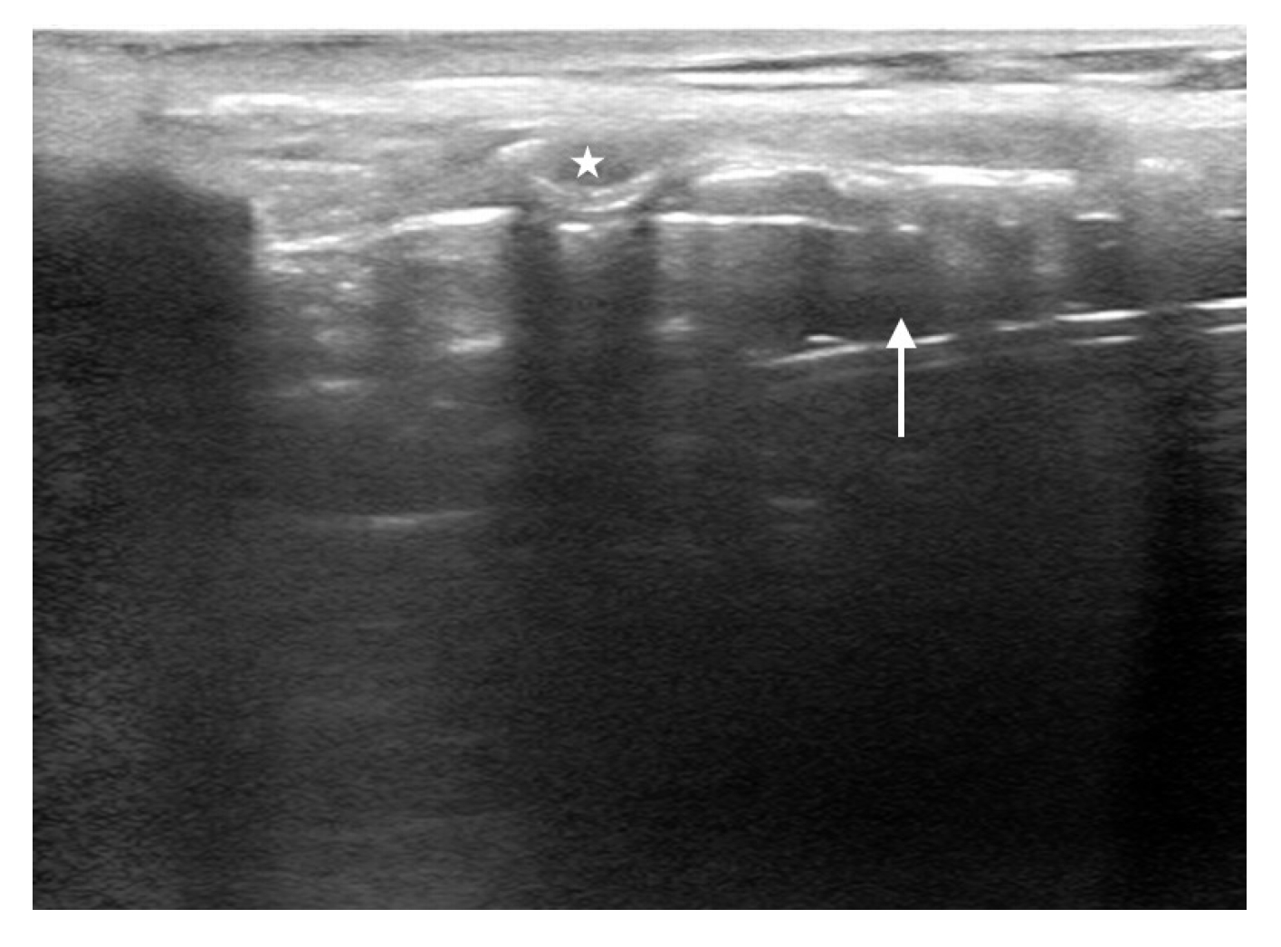
The technique to identify the CTM is similar to ETT depth determination described above. Lay the patient supine with their neck extended. Start with the transducer in the sagittal plane on the anterior neck just inferior to the thyroid cartilage. Identify the tracheal rings, which will appear as a “string of pearls” leading from caudal to cranial direction. Immediately superior to that will be a larger ring (i.e., cricoid cartilage), followed by a hyperechoic membrane and then a larger rectangular structure (i.e., thyroid cartilage). The hyperechoic line between and just deep to the cricoid and thyroid cartilages is the CTM (Fig. 9). Slide a blunt needle under the cranial portion of the transducer down to the corresponding location of the CTM and subsequently mark the skin with a surgical pen [84].
Best practice is to mark the CTM prior to the intubation, rather than attempting to identify it after a failed intubation attempt. The CTM should be marked in the position that the cricothyrotomy would be performed. One study [96] found that the midpoint of the CTM moved caudally an average of 4.2 mm when changing the head of bed elevation from 90° to 0° and this effect was increased if the patient was obese or over age 70 years [96]. Moreover, when the CTM is marked in the neutral neck position, the marking can move outside the CTM border when the neck is converted to full extension [97,98]. Fortunately, the CTM will return to the correct location when the patient is returned to the same position as when they were originally marked [99]. Therefore, it is advisable to mark the CTM in neck extension as close to the anticipated procedural position as possible prior to any attempted airway intervention [84,100].

ULTRASOUND FOR THE CRASHING VENTILATED PATIENT

Ultrasound also offers benefits in the assessment of the ventilated patient who experiences clinical deterioration. Traditionally, assessment of the postintubation patient has relied primarily on indirect assessment using structured algorithms, such as DOPES (dislodgement, obstruction, pneumothorax, equipment malfunction, stacking of breaths). However, as POCUS has expanded, we propose an update algorithm: Sono-DOPES. This algorithm builds upon previous work in DOPES (which had relied upon primarily physical examination findings) and adds ultrasound to enhance each stage (Table 1).

CONCLUSION

Airway management is a common procedure within emergency and critical care medicine. Traditional techniques for predicting and managing a difficult airway each have important limitations. As the field has evolved, POCUS has been increasingly utilized for this application. Ultrasound can be utilized to predict difficult airways, confirm ETT location and depth, locate the CTM, and facilitate the assessment and management of the crashing patient on mechanical ventilation.

NOTES

Author contributions
Conceptualization: all authors; Supervision: MG; Writing–original draft: all authors; Writing–review & editing: all authors. All authors read and approved the final manuscript.
Conflicts of interest
The authors have no conflicts of interest to declare.
Funding
The authors received no financial support for this study.
Data availability
Data sharing is not applicable as no new data were created or analyzed in this study.

REFERENCES

1. Cairns C, Kang K; National Center for Health Statistics. National Hospital Ambulatory Medical Care Survey: 2020 emergency department summary tables. Centers for Disease Control and Prevention (CDC); Prevention; 2022..
crossref
2. Turner JS, Bucca AW, Propst SL, et al. Association of checklist use in endotracheal intubation with clinically important outcomes: a systematic review and meta-analysis. JAMA Netw Open 2020; 3:e209278.
crossref pmid pmc
3. De Jong A, Rolle A, Molinari N, et al. Cardiac arrest and mortality related to intubation procedure in critically ill adult patients: a multicenter cohort study. Crit Care Med 2018; 46:532-9.
crossref pmid
4. Brown CA 3rd, Bair AE, Pallin DJ, Walls RM; NEAR III Investigators. Techniques, success, and adverse events of emergency department adult intubations. Ann Emerg Med 2015; 65:363-70.
crossref pmid
5. Gottlieb M, Cosby K. Ultrasound-guided hematoma block for distal radial and ulnar fractures. J Emerg Med 2015; 48:310-2.
crossref pmid
6. Gottlieb M, Sundaram T, Holladay D, Nakitende D. Ultrasound-guided peripheral intravenous line placement: a narrative review of evidence-based best practices. West J Emerg Med 2017; 18:1047-54.
crossref pmid pmc
7. Gottlieb M, Holladay D, Peksa GD. Ultrasound-assisted lumbar punctures: a systematic review and meta-analysis. Acad Emerg Med 2019; 26:85-96.
crossref pmid pdf
8. Gottlieb M, Patel D, Marks A, Peksa GD. Ultrasound for the diagnosis of shoulder dislocation and reduction: a systematic review and meta-analysis. Acad Emerg Med 2022; 29:999-1007.
pmid
9. Gottlieb M, Alerhand S. Managing cardiac arrest using ultrasound. Ann Emerg Med 2023; 81:532-42.
crossref pmid
10. Gottlieb M, Holladay D, Peksa GD. Ultrasonography for the confirmation of endotracheal tube intubation: a systematic review and meta-analysis. Ann Emerg Med 2018; 72:627-36.
crossref pmid
11. Gottlieb M, Olszynski P, Atkinson P. Just the facts: point-of-care ultrasound for airway management. CJEM 2021; 23:277-9.
crossref pmid pdf
12. Norskov AK, Rosenstock CV, Wetterslev J, Astrup G, Afshari A, Lundstrom LH. Diagnostic accuracy of anaesthesiologists’ prediction of difficult airway management in daily clinical practice: a cohort study of 188 064 patients registered in the Danish Anaesthesia Database. Anaesthesia 2015; 70:272-81.
crossref pmid pdf
13. Long B, Koyfman A, Gottlieb M. Factors predicting difficult endotracheal intubation. Acad Emerg Med 2019; 26:1294-6.
crossref pmid pdf
14. Karamchandani K, Wheelwright J, Yang AL, Westphal ND, Khanna AK, Myatra SN. Emergency airway management outside the operating room: current evidence and management strategies. Anesth Analg 2021; 133:648-62.
crossref pmid
15. Detsky ME, Jivraj N, Adhikari NK, et al. Will this patient be difficult to intubate?: the rational clinical examination systematic review. JAMA 2019; 321:493-503.
crossref pmid
16. Roth D, Pace NL, Lee A, et al. Airway physical examination tests for detection of difficult airway management in apparently normal adult patients. Cochrane Database Syst Rev 2018; 5:CD008874.
crossref pmid
17. Adhikari S, Zeger W, Schmier C, et al. Pilot study to determine the utility of point-of-care ultrasound in the assessment of difficult laryngoscopy. Acad Emerg Med 2011; 18:754-8.
crossref pmid
18. Pinto J, Cordeiro L, Pereira C, Gama R, Fernandes HL, Assuncao J. Predicting difficult laryngoscopy using ultrasound measurement of distance from skin to epiglottis. J Crit Care 2016; 33:26-31.
crossref pmid
19. Nakazawa H, Uzawa K, Tokumine J, Lefor AK, Motoyasu A, Yorozu T. Airway ultrasound for patients anticipated to have a difficult airway: perspective for personalized medicine. World J Clin Cases 2023; 11:1951-62.
crossref pmid pmc
20. Gottlieb M, Holladay D, Burns KM, Nakitende D, Bailitz J. Ultrasound for airway management: an evidence-based review for the emergency clinician. Am J Emerg Med 2020; 38:1007-13.
crossref pmid
21. Lin J, Bellinger R, Shedd A, et al. Point-of-care ultrasound in airway evaluation and management: a comprehensive review. Diagnostics (Basel) 2023; 13:1541.
crossref pmid pmc
22. Carsetti A, Sorbello M, Adrario E, Donati A, Falcetta S. Airway ultrasound as predictor of difficult direct laryngoscopy: a systematic review and meta-analysis. Anesth Analg 2022; 134:740-50.
crossref pmid pmc
23. Gomes SH, Simoes AM, Nunes AM, et al. Useful ultrasonographic parameters to predict difficult laryngoscopy and difficult tracheal intubation: a systematic review and meta-analysis. Front Med (Lausanne) 2021; 8:671658.
crossref pmid pmc
24. Komatsu R, Sengupta P, Wadhwa A, et al. Ultrasound quantification of anterior soft tissue thickness fails to predict difficult laryngoscopy in obese patients. Anaesth Intensive Care 2007; 35:32-7.
crossref pdf
25. Andruszkiewicz P, Wojtczak J, Sobczyk D, Stach O, Kowalik I. Effectiveness and validity of sonographic upper airway evaluation to predict difficult laryngoscopy. J Ultrasound Med 2016; 35:2243-52.
crossref pmid pdf
26. Wu H, Wang H. Diagnostic efficacy and clinical value of ultrasonography in difficult airway assessment: based on a prospective cohort study. Contrast Media Mol Imaging 2022; 2022:4706438.
crossref pmid pmc pdf
27. Petrisor C, Szabo R, Constantinescu C, Prie A, Hagau N. Ultrasound-based assessment of hyomental distances in neutral, ramped, and maximum hyperextended positions, and derived ratios, for the prediction of difficult airway in the obese population: a pilot diagnostic accuracy study. Anaesthesiol Intensive Ther 2018; 50:110-6.
crossref pmid
28. Yadav NK, Rudingwa P, Mishra SK, Pannerselvam S. Ultrasound measurement of anterior neck soft tissue and tongue thickness to predict difficult laryngoscopy: an observational analytical study. Indian J Anaesth 2019; 63:629-34.
crossref pmid pmc
29. Yao W, Wang B. Can tongue thickness measured by ultrasonography predict difficult tracheal intubation? Br J Anaesth 2017; 118:601-9.
crossref pmid
30. Park L, Zeng I, Brainard A. Systematic review and meta-analysis of first-pass success rates in emergency department intubation: creating a benchmark for emergency airway care. Emerg Med Australas 2017; 29:40-7.
crossref pmid pdf
31. Crewdson K, Lockey DJ, Roislien J, Lossius HM, Rehn M. The success of pre-hospital tracheal intubation by different pre-hospital providers: a systematic literature review and meta-analysis. Crit Care 2017; 21:31.
crossref pmid pmc pdf
32. Kelly JJ, Eynon CA, Kaplan JL, de Garavilla L, Dalsey WC. Use of tube condensation as an indicator of endotracheal tube placement. Ann Emerg Med 1998; 31:575-8.
crossref pmid
33. Tanigawa K, Takeda T, Goto E, Tanaka K. Accuracy and reliability of the self-inflating bulb to verify tracheal intubation in out-of-hospital cardiac arrest patients. Anesthesiology 2000; 93:1432-6.
crossref pmid pdf
34. Tanigawa K, Takeda T, Goto E, Tanaka K. The efficacy of esophageal detector devices in verifying tracheal tube placement: a randomized cross-over study of out-of-hospital cardiac arrest patients. Anesth Analg 2001; 92:375-8.
crossref pmid
35. Takeda T, Tanigawa K, Tanaka H, Hayashi Y, Goto E, Tanaka K. The assessment of three methods to verify tracheal tube placement in the emergency setting. Resuscitation 2003; 56:153-7.
crossref pmid
36. Grmec S. Comparison of three different methods to confirm tracheal tube placement in emergency intubation. Intensive Care Med 2002; 28:701-4.
crossref pmid pdf
37. Roy PS, Joshi N, Garg M, Meena R, Bhati S. Comparison of ultrasonography, clinical method and capnography for detecting correct endotracheal tube placement: a prospective, observational study. Indian J Anaesth 2022; 66:826-31.
crossref pmid pmc
38. Li J. Capnography alone is imperfect for endotracheal tube placement confirmation during emergency intubation. J Emerg Med 2001; 20:223-9.
crossref pmid
39. Eichlseder M, Eichinger M, Pichler A, et al. Out-of-hospital arterial to end-tidal carbon dioxide gradient in patients with return of spontaneous circulation after out-of-hospital cardiac arrest: a retrospective study. Ann Emerg Med 2023; 82:558-63.
crossref
40. Chenkin J, McCartney CJ, Jelic T, Romano M, Heslop C, Bandiera G. Defining the learning curve of point-of-care ultrasound for confirming endotracheal tube placement by emergency physicians. Crit Ultrasound J 2015; 7:14.
crossref pmid pmc pdf
41. Long B, Koyfman A, Gottlieb M. Diagnostic accuracy of ultrasound for confirmation of endotracheal tube placement. Acad Emerg Med 2019; 26:1096-8.
crossref pmid pdf
42. Lin MJ, Gurley K, Hoffmann B. Bedside ultrasound for tracheal tube verification in pediatric emergency department and ICU patients: a systematic review. Pediatr Crit Care Med 2016; 17:e469-76.
crossref pmid
43. Gottlieb M, Holladay D, Nakitende D, et al. Variation in the accuracy of ultrasound for the detection of intubation by endotracheal tube size. Am J Emerg Med 2019; 37:706-9.
crossref pmid
44. Gottlieb M, Holladay D, Burns K, et al. Accuracy of ultrasound for endotracheal intubation between different transducer types. Am J Emerg Med 2019; 37:2182-5.
crossref pmid
45. Lonchena T, So S, Ibinson J, Roolf P, Orebaugh SL. Optimization of ultrasound transducer positioning for endotracheal tube placement confirmation in cadaveric model. J Ultrasound Med 2017; 36:279-84.
crossref pmid pdf
46. Romano MJ, Lee JS, Chenkin J. Comparison of techniques for visualisation of the airway anatomy for ultrasound-assisted intubation: a prospective study of emergency department patients. Anaesth Crit Care Pain Med 2018; 37:545-9.
crossref pmid
47. Gottlieb M, Nakitende D, Sundaram T, Serici A, Shah S, Bailitz J. Comparison of static versus dynamic ultrasound for the detection of endotracheal intubation. West J Emerg Med 2018; 19:412-6.
crossref pmid pmc
48. Gottlieb M, Patel D, Sundaram T. Comparison of endotracheal tube location on ultrasound accuracy, time, and confidence. Am J Emerg Med 2022; 62:127-8.
crossref pmid
49. Gottlieb M, Burns K, Holladay D, Chottiner M, Shah S, Gore SR. Impact of endotracheal tube twisting on the diagnostic accuracy of ultrasound for intubation confirmation. Am J Emerg Med 2020; 38:1332-4.
crossref pmid
50. Gottlieb M, Holladay D, Serici A, Shah S, Nakitende D. Comparison of color flow with standard ultrasound for the detection of endotracheal intubation. Am J Emerg Med 2018; 36:1166-9.
crossref pmid
51. Uya A, Spear D, Patel K, Okada P, Sheeran P, McCreight A. Can novice sonographers accurately locate an endotracheal tube with a saline-filled cuff in a cadaver model? A pilot study. Acad Emerg Med 2012; 19:361-4.
crossref pmid
52. Tessaro MO, Arroyo AC, Haines LE, Dickman E. Inflating the endotracheal tube cuff with saline to confirm correct depth using bedside ultrasonography. CJEM 2015; 17:94-8.
crossref pmid
53. Gottlieb M, Patel D, Jung C, et al. Comparison of saline versus air for identifying endotracheal intubation with ultrasound. Am J Emerg Med 2022; 58:131-4.
crossref pmid
54. Weaver B, Lyon M, Blaivas M. Confirmation of endotracheal tube placement after intubation using the ultrasound sliding lung sign. Acad Emerg Med 2006; 13:239-44.
crossref pmid
55. Sim SS, Lien WC, Chou HC, et al. Ultrasonographic lung sliding sign in confirming proper endotracheal intubation during emergency intubation. Resuscitation 2012; 83:307-12.
crossref pmid
56. Park SC, Ryu JH, Yeom SR, Jeong JW, Cho SJ. Confirmation of endotracheal intubation by combined ultrasonographic methods in the emergency department. Emerg Med Australas 2009; 21:293-7.
crossref pmid
57. Saglam C, Unluer EE, Karagoz A. Confirmation of endotracheal tube position during resuscitation by bedside ultrasonography. Am J Emerg Med 2013; 31:248-50.
crossref pmid
58. Abbasi S, Farsi D, Zare MA, Hajimohammadi M, Rezai M, Hafezimoghadam P. Direct ultrasound methods: a confirmatory technique for proper endotracheal intubation in the emergency department. Eur J Emerg Med 2015; 22:10-6.
pmid
59. Rajan S, Surendran J, Paul J, Kumar L. Rapidity and efficacy of ultrasonographic sliding lung sign and auscultation in confirming endotracheal intubation in overweight and obese patients. Indian J Anaesth 2017; 61:230-4.
crossref pmid pmc
60. Hsieh KS, Lee CL, Lin CC, Huang TC, Weng KP, Lu WH. Secondary confirmation of endotracheal tube position by ultrasound image. Crit Care Med 2004; 32(9 Suppl):S374-7.
crossref pmid
61. Kerrey BT, Geis GL, Quinn AM, Hornung RW, Ruddy RM. A prospective comparison of diaphragmatic ultrasound and chest radiography to determine endotracheal tube position in a pediatric emergency department. Pediatrics 2009; 123:e1039-44.
crossref pmid pdf
62. Hosseini JS, Talebian MT, Ghafari MH, Eslami V. Secondary confirmation of endotracheal tube position by diaphragm motion in right subcostal ultrasound view. Int J Crit Illn Inj Sci 2013; 3:113-7.
crossref pmid pmc
63. Cavo JW Jr. True vocal cord paralysis following intubation. Laryngoscope 1985; 95:1352-9.
crossref pmid
64. Conrardy PA, Goodman LR, Lainge F, Singer MM. Alteration of endotracheal tube position. Flexion and extension of the neck. Crit Care Med 1976; 4:8-12.
crossref pmid
65. Alerhand S, Tsung JW. Unmasking the lung pulse for detection of endobronchial intubation. J Ultrasound Med 2020; 39:2105-9.
crossref pmid pdf
66. Owen RL, Cheney FW. Endobronchial intubation: a preventable complication. Anesthesiology 1987; 67:255-7.
pmid
67. Men XQ, Yan XX. Tracheal ultrasound for the accurate confirmation of the endotracheal tube position in obese patients. J Ultrasound Med 2020; 39:509-13.
crossref pmid pdf
68. Harris EA, Arheart KL, Penning DH. Endotracheal tube malposition within the pediatric population: a common event despite clinical evidence of correct placement. Can J Anaesth 2008; 55:685-90.
pmid
69. Brunel W, Coleman DL, Schwartz DE, Peper E, Cohen NH. Assessment of routine chest roentgenograms and the physical examination to confirm endotracheal tube position. Chest 1989; 96:1043-5.
crossref pmid
70. Sitzwohl C, Langheinrich A, Schober A, et al. Endobronchial intubation detected by insertion depth of endotracheal tube, bilateral auscultation, or observation of chest movements: randomised trial. BMJ 2010; 341:c5943.
crossref pmid pmc
71. Parab SY, Kumar P, Divatia JV, Sharma K. A prospective randomized controlled double-blind study comparing auscultation and lung ultrasonography in the assessment of double lumen tube position in elective thoracic surgeries involving one lung ventilation at a tertiary care cancer institute. Korean J Anesthesiol 2019; 72:24-31.
crossref pmid pdf
72. Hossein-Nejad H, Payandemehr P, Bashiri SA, Nedai HH. Chest radiography after endotracheal tube placement: is it necessary or not? Am J Emerg Med 2013; 31:1181-2.
crossref pmid
73. Gottlieb M, Berzins D, Hartrich M, et al. Diagnostic accuracy of ultrasound to confirm endotracheal tube depth. Am J Emerg Med 2022; 62:9-13.
crossref pmid
74. Yang FM, Ma BZ, Liu Y, et al. Lung ultrasound for detecting tracheal and mainstem intubation: a systematic review and meta-analysis. Ultrasound Med Biol 2022; 48:3-9.
crossref pmid
75. Mori T, Nomura O, Hagiwara Y, Inoue N. Diagnostic accuracy of a 3-point ultrasound protocol to detect esophageal or endobronchial mainstem intubation in a pediatric emergency department. J Ultrasound Med 2019; 38:2945-54.
crossref pmid pdf
76. Uya A, Gautam NK, Rafique MB, et al. Point-of-care ultrasound in sternal notch confirms depth of endotracheal tube in children. Pediatr Crit Care Med 2020; 21:e393-8.
crossref pmid
77. Lichtenstein DA, Lascols N, Prin S, Meziere G. The “lung pulse”: an early ultrasound sign of complete atelectasis. Intensive Care Med 2003; 29:2187-92.
crossref pmid pdf
78. Gottlieb M, Alerhand S, Long B. Point-of-care ultrasound for intubation confirmation of COVID-19 patients. West J Emerg Med 2020; 21:1042-5.
crossref pmid pmc
79. Morray JP, Geiduschek JM, Caplan RA, Posner KL, Gild WM, Cheney FW. A comparison of pediatric and adult anesthesia closed malpractice claims. Anesthesiology 1993; 78:461-7.
crossref pmid pdf
80. Peterson GN, Domino KB, Caplan RA, Posner KL, Lee LA, Cheney FW. Management of the difficult airway: a closed claims analysis. Anesthesiology 2005; 103:33-9.
pmid
81. Cook TM, MacDougall-Davis SR. Complications and failure of airway management. Br J Anaesth 2012; 109 Suppl 1:i68-85.
crossref pmid
82. Melchiors J, Todsen T, Nilsson P, et al. Preparing for emergency: a valid, reliable assessment tool for emergency cricothyroidotomy skills. Otolaryngol Head Neck Surg 2015; 152:260-5.
pmid
83. Cho SA, Kang P, Song IS, et al. Performance time of anesthesiology trainees for cricothyroid membrane identification and characteristics of cricothyroid membrane in pediatric patients using ultrasonography. Paediatr Anaesth 2022; 32:834-42.
crossref pmid pdf
84. Kristensen MS, Teoh WH. Ultrasound identification of the cricothyroid membrane: the new standard in preparing for front-of-neck airway access. Br J Anaesth 2021; 126:22-7.
crossref pmid
85. Cook TM, Woodall N, Frerk C; Fourth National Audit Project. Major complications of airway management in the UK: results of the Fourth National Audit Project of the Royal College of Anaesthetists and the Difficult Airway Society. Part 1: anaesthesia. Br J Anaesth 2011; 106:617-31.
crossref pmid
86. You-Ten KE, Desai D, Postonogova T, Siddiqui N. Accuracy of conventional digital palpation and ultrasound of the cricothyroid membrane in obese women in labour. Anaesthesia 2015; 70:1230-4.
crossref pmid
87. You-Ten KE, Wong DT, Ye XY, Arzola C, Zand A, Siddiqui N. Practice of ultrasound-guided palpation of neck landmarks improves accuracy of external palpation of the cricothyroid membrane. Anesth Analg 2018; 127:1377-82.
crossref pmid
88. Siddiqui N, Yu E, Boulis S, You-Ten KE. Ultrasound is superior to palpation in identifying the cricothyroid membrane in subjects with poorly defined neck landmarks: a randomized clinical trial. Anesthesiology 2018; 129:1132-9.
pmid
89. Siddiqui N, Arzola C, Friedman Z, Guerina L, You-Ten KE. Ultrasound improves cricothyrotomy success in cadavers with poorly defined neck anatomy: a randomized control trial. Anesthesiology 2015; 123:1033-41.
pmid
90. Mandell D, Orebaugh SL. A porcine model for learning ultrasound anatomy of the larynx and ultrasound-guided cricothyrotomy. Simul Healthc 2019; 14:343-7.
crossref pmid
91. Acuna J, Pacheco G, Yarnish AA, et al. A novel simulation model for training emergency medicine residents in the ultrasound identification of landmarks for cricothyrotomy. Cureus 2022; 14:e33003.
pmid pmc
92. Oliveira KF, Arzola C, Ye XY, Clivatti J, Siddiqui N, You-Ten KE. Determining the amount of training needed for competency of anesthesia trainees in ultrasonographic identification of the cricothyroid membrane. BMC Anesthesiol 2017; 17:74.
crossref pmid pmc pdf
93. Alerhand S. Ultrasound for identifying the cricothyroid membrane prior to the anticipated difficult airway. Am J Emerg Med 2018; 36:2078-84.
crossref pmid
94. Lavelle A, Drew T, Fennessy P, McCaul C, Shannon J. Accuracy of cricothyroid membrane identification using ultrasound and palpation techniques in obese obstetric patients: an observational study. Int J Obstet Anesth 2021; 48:103205.
crossref pmid
95. Hung KC, Chen IW, Lin CM, Sun CK. Comparison between ultrasound-guided and digital palpation techniques for identification of the cricothyroid membrane: a meta-analysis. Br J Anaesth 2021; 126:e9-11.
crossref pmid
96. Arthurs L, Erdelyi S, Kim DJ. The effect of patient positioning on ultrasound landmarking for cricothyrotomy. Can J Anaesth 2021; 68:24-9.
crossref pmid pdf
97. Dixit A, Ramaswamy KK, Perera S, Sukumar V, Frerk C. Impact of change in head and neck position on ultrasound localisation of the cricothyroid membrane: an observational study. Anaesthesia 2019; 74:29-32.
crossref pmid pdf
98. Fennessy P, Drew T, Husarova V, Duggan M, McCaul CL. Emergency cricothyroidotomy: an observational study to estimate optimal incision position and length. Br J Anaesth 2019; 122:263-8.
crossref pmid
99. Bowness J, Teoh WH, Kristensen MS, et al. A marking of the cricothyroid membrane with extended neck returns to correct position after neck manipulation and repositioning. Acta Anaesthesiol Scand 2020; 64:1422-5.
crossref pmid pdf
100. Rai Y, You-Ten E, Zasso F, De Castro C, Ye XY, Siddiqui N. The role of ultrasound in front-of-neck access for cricothyroid membrane identification: a systematic review. J Crit Care 2020; 60:161-8.
crossref pmid

Fig. 1.
Distance from the skin to the epiglottis. Example measurement from skin to anterior aspect of the epiglottis (star).
ceem-23-094f1.jpg
Fig. 2.
Distance from the skin to the vocal cords. Example measurement from skin to anterior aspect of the vocal cords. The arrow indicates the anterior vocal cord commissure.
ceem-23-094f2.jpg
Fig. 3.
Hyomental distance. Example measurement of hyomental distance. The arrow indicates the mentum. The star indicates the hyoid.
ceem-23-094f3.jpg
Fig. 4.
Tongue thickness. Example measurement from skin to posterior aspect of the tongue (star).
ceem-23-094f4.jpg
Fig. 5.
Difficult Airway Evaluation with Sonography (DARES) algorithm. DSE, distance from the skin to the epiglottis; HMD, hyomental distance; TT, tongue thickness; HMDR2, ratio of the HMD measured with the head in maximal extension and the head in neutral position; HMDR1, ratio of the HMD measured with the head in a ramped position and the head in neutral position. Adapted from Lin et al. [21], available under the Creative Commons License.
ceem-23-094f5.jpg
Fig. 6.
Tracheal intubation. The arrow indicates the anterior aspect of endotracheal tube. T, trachea.
ceem-23-094f6.jpg
Fig. 7.
Esophageal intubation. In the case of esophageal intubation, a curvilinear structure which mimics the trachea (T), is seen to the right of the trachea. This is the endotracheal tube within the esophagus (E).
ceem-23-094f7.jpg
Fig. 8.
Endotracheal tube in the correct depth. The star indicates the cricoid cartilage. The arrow indicates the endotracheal tube cuff.
ceem-23-094f8.jpg
Fig. 9.
Cricothyroid membrane. The arrowhead indicates the thyroid cartilage. The arrow indicates the cricothyroid membrane. The star indicates the cricoid cartilage.
ceem-23-094f9.jpg
Table 1.
Sono-DOPES for the crashing ventilated patient
Potential etiology Diagnostic assessment
Dislodgment Transtracheal ultrasound to assess for endotracheal vs. esophageal location and ETT depth
Obstruction of ETT Lung ultrasound to assess for lung sliding to demonstrate air is entering the lungs through the ETT
Pneumothorax Lung ultrasound to assess for bilateral lung sliding
If unilateral lung sliding is present, lung ultrasound assess for lung pulse to differentiate single lung intubation versus pneumothorax
Equipment malfunction Lung ultrasound to assess for lung sliding
If no lung sliding and ETT is in the appropriate location, disconnect from the ventilator and repeat lung ultrasound with manual ventilation
Stacking of breaths Lung ultrasound to assess for bilateral lung sliding
Diaphragm ultrasound to assess for diaphragmatic expansion to suggest adequate air movement

DOPES, dislodgement, obstruction, pneumothorax, equipment malfunction, stacking of breaths; ETT, endotracheal tube.

Editorial Office
The Korean Society of Emergency Medicine
101-3104, Brownstone Seoul, 464 Cheongpa-ro, Jung-gu, Seoul 04510, Korea
TEL: +82-31-709-0918   E-mail: office@ceemjournal.org
About |  Browse Articles |  Current Issue |  For Authors and Reviewers
Copyright © by The Korean Society of Emergency Medicine.                 Developed in M2PI