| Home | E-Submission | Sitemap | Contact Us |  
Search
Clin Exp Emerg Med Search

CLOSE

Clin Exp Emerg Med > Volume 10(3); 2023 > Article
Shalaby and Sahni: Supraclavicular brachial plexus block: the unsung hero of emergency department regional anesthesia
Dear Editor,
In the mid-2000s, the supraclavicular brachial plexus (SBP) block surfaced in emergency medicine literature; case reports and series demonstrated it as an effective means for managing challenging upper extremity (UE) injuries [14]. Despite its appeal, the SBP block has not gained significant traction in emergency departments (EDs) and is taught rarely in academic emergency medicine curricula. We believe that, though the SBP block is underappreciated, it is extremely valuable and should be learned, taught, and utilized in emergency practice.
One of the main advantages of SBP block is that it is simple to setup. Most brachial plexus blocks involve special equipment not readily accessible in the ED, such as spinal needles, intravenous tubing, and a long linear probe for ultrasound (US). An SBP block, however, only requires a 22-gauge needle, a 20-mL syringe, and a simple short linear probe already included on most US systems.
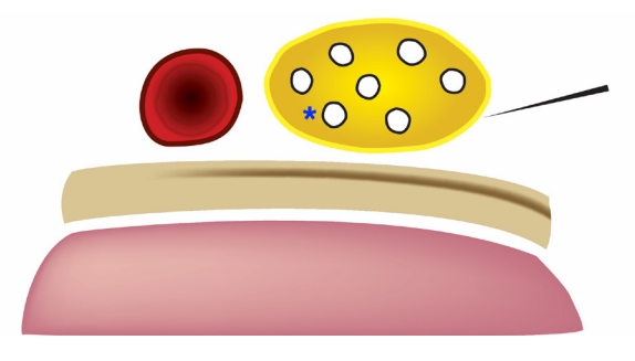
During the procedure, patients can position the UE in adduction as comfortably as possible since no special positioning is required for the procedure. In addition, the SBP block is simple to learn and perform. With the linear probe situated in the supraclavicular fossa, the SBP is visualized on US immediately lateral to the subclavian artery and superior to the first rib and pleura. The shallow position of 1 to 2 cm below the skin surface allows easy accessibility to the SBP [5]. Under US guidance, the physician directs a needle in-plane from lateral to medial toward the SBP and instills local anesthetic within or just outside the sheath (Supplementary Video 1). Injecting at the inferior “corner pocket” nearest the subclavian artery within the SBP sheath guarantees dense anesthesia directly targeting the inferior trunk (Figs. 1, 2) [6,7]. The onset of anesthesia occurs within minutes of injection [6].
To complement its easy learning curve, the SBP block boasts an excellent safety profile. Though pneumothorax is a well-known complication, US guidance has significantly minimized this risk [811]. Also, the first rib acts as a backstop to the needle path and may prevent pneumothorax even if the physician advances past the SBP. As with all brachial plexus blocks above the clavicle, hemidiaphragmatic paralysis may persist in up to 70% of patients after an SBP block [12]. However, Renes et al. [13] demonstrated significantly lower rates of hemidiaphragmatic paralysis using less than 20 mL of ropivacaine for a SBP block. Transient hemidiaphragmatic paralysis secondary to phrenic nerve palsy is common after brachial plexus blocks such as the interscalene nerve block, SBP block, and infraclavicular brachial plexus block [14,15]. Yet healthy patients without chronic cardiac or pulmonary disease or obesity tend not to experience respiratory compromise [14], perhaps because the minor respiratory muscles (sternocleidomastoid, scalene, and intercostals) and contralateral hemidiaphragm compensate. While vascular puncture is also a concern, the subclavian artery lies on the opposite side of the approach, and there are no major arteries in the needle trajectory. Additionally, neuropraxia is a worrisome but relatively infrequent outcome [16]. In a retrospective review performed by Liu et al. [17], 17% of perioperative US-guided SBP blocks involved inadvertent intraneural injection, but no patients suffered brachial plexus injury. Other complications include Horner syndrome and, as with any nerve block, local anesthetic systemic toxicity (LAST). Similar to hemidiaphragmatic paralysis, the risk of LAST can be minimized with lower doses of anesthetic [18].
The SBP block is a safe and powerful technique that enables physicians to anesthetize the entire upper limb including the shoulder in the frequent UE injuries presenting to the ED [5]. There is no equivalent nerve block for the chest, back, or lower extremities. In our ED, for example, we have employed the SBP block for anterior shoulder dislocations; wrist, elbow, and humerus fractures; complicated lacerations; and postoperative compressive neuropathy. Published reports have used it for UE abscesses, fractures, and joint dislocations [14]. While other brachial plexus blocks are useful for UE anesthesia, each has its own imperfections. The interscalene block, for example, only anesthetizes the sensory distribution of C5–C7 and excludes the inferior trunk (C8–T1) of the brachial plexus, limiting its use for injuries distal to the elbow. The infraclavicular brachial plexus block, on the other hand, involves a daunting needle trajectory aimed toward the pleura and may also require multiple needle passes. The blind needle path of the retroclavicular approach to the infraclavicular region under the clavicle is followed by a narrow lane between the pleura and the axillary artery, hindering novice users. The axillary nerve block also involves multiple needle passes and requires that the patient abduct the arm, making its impractical for particular injuries.
Musculoskeletal pain is the most common complaint in the ED, and there is no one-size-fits-all technique for management. While the SBP block is not superior to all other brachial plexus blocks, it is undervalued and underutilized in emergency medicine. Considering the ease, safety, and potency of the SBP block, emergency physicians should include the SBP block in the multimodal approach to acute pain.

SUPPLEMENTARY MATERIAL

Supplementary material is available at https://doi.org/10.15441/ceem.23.035.

Supplemental Video 1.

Performance of a left-sided supraclavicular brachial plexus block. The left side of the screen is medial; the right side is lateral. With a high-frequency linear probe oriented obliquely in the supraclavicular fossa, the supraclavicular brachial plexus is immediately lateral to the subclavian artery.

NOTES

ETHICS STATEMENTS
Not applicable.
CONFLICT OF INTEREST
No potential conflict of interest relevant to this article was reported.
FUNDING
None.
AUTHOR CONTRIBUTIONS
Conceptualization: all authors; Project administration: MS; Resources: MS; Supervision: MS; Visualization: all authors; Writing–original draft: all authors; Writing–review & editing: all authors. All authors read and approved the final manuscript.

REFERENCES

1. Stone MB, Wang R, Price DD. Ultrasound-guided supraclavicular brachial plexus nerve block vs procedural sedation for the treatment of upper extremity emergencies. Am J Emerg Med 2008; 26:706-10.
crossref pmid
2. Bhoi S, Chandra A, Galwankar S. Ultrasound-guided nerve blocks in the emergency department. J Emerg Trauma Shock 2010; 3:82-8.
crossref pmid pmc
3. Shahar-Nissan K, Berant R, Ganor L, Katzir Y. Ultrasound-guided supraclavicular brachial plexus blocks performed by pediatric emergency medicine physicians for painful orthopedic procedures in a pediatric emergency department: a case series. Pediatr Emerg Care 2022; 38:e1684-7.
crossref pmid
4. Stone MB, Price DD, Wang R. Ultrasound-guided supraclavicular block for the treatment of upper extremity fractures, dislocations, and abscesses in the ED. Am J Emerg Med 2007; 25:472-5.
crossref pmid
6. Soares LG, Brull R, Lai J, Chan VW. Eight ball, corner pocket: the optimal needle position for ultrasound-guided supraclavicular block. Reg Anesth Pain Med 2007; 32:94-5.
crossref pmid
7. Shalaby M, Smith M, Tran L, Farrow R. Utility of supraclavicular brachial plexus block for anterior shoulder dislocation: could it be iseful? West J Emerg Med 2023; 24:793-7.
pmid pmc
8. Perlas A, Lobo G, Lo N, Brull R, Chan VW, Karkhanis R. Ultrasound-guided supraclavicular block: outcome of 510 consecutive cases. Reg Anesth Pain Med 2009; 34:171-6.
pmid
9. Williams SR, Chouinard P, Arcand G, et al. Ultrasound guidance speeds execution and improves the quality of supraclavicular block. Anesth Analg 2003; 97:1518-23.
crossref pmid
10. Kapral S, Krafft P, Eibenberger K, Fitzgerald R, Gosch M, Weinstabl C. Ultrasound-guided supraclavicular approach for regional anesthesia of the brachial plexus. Anesth Analg 1994; 78:507-13.
crossref pmid
11. Franco CD, Gloss FJ, Voronov G, Tyler SG, Stojiljkovic LS. Supraclavicular block in the obese population: an analysis of 2020 blocks. Anesth Analg 2006; 102:1252-4.
crossref pmid
12. Hong B, Lee S, Oh C, et al. Hemidiaphragmatic paralysis following costoclavicular versus supraclavicular brachial plexus block: a randomized controlled trial. Sci Rep 2021; 11:18749.
crossref pmid pmc pdf
13. Renes SH, Spoormans HH, Gielen MJ, Rettig HC, van Geffen GJ. Hemidiaphragmatic paresis can be avoided in ultrasound-guided supraclavicular brachial plexus block. Reg Anesth Pain Med 2009; 34:595-9.
crossref pmid
14. Bergmann L, Martini S, Kesselmeier M, et al. Phrenic nerve block caused by interscalene brachial plexus block: breathing effects of different sites of injection. BMC Anesthesiol 2016; 16:45.
crossref pmid pmc pdf
15. Petrar SD, Seltenrich ME, Head SJ, Schwarz SK. Hemidiaphragmatic paralysis following ultrasound-guided supraclavicular versus infraclavicular brachial plexus blockade: a randomized clinical trial. Reg Anesth Pain Med 2015; 40:133-8.
pmid
16. Gadsden J, Orebaugh S. Targeted intracluster supraclavicular brachial plexus block: too close for comfort. Br J Anaesth 2019; 122:713-5.
crossref pmid
17. Liu SS, YaDeau JT, Shaw PM, Wilfred S, Shetty T, Gordon M. Incidence of unintentional intraneural injection and postoperative neurological complications with ultrasound-guided interscalene and supraclavicular nerve blocks. Anaesthesia 2011; 66:168-74.
crossref pmid
18. Dickerson DM, Apfelbaum JL. Local anesthetic systemic toxicity. Aesthet Surg J 2014; 34:1111-9.
crossref pmid

Fig. 1.
Supraclavicular brachial plexus (yellow), subclavian artery (red), first rib (beige), and pleura (pink). The needle (upper right corner of the image) approaches the supraclavicular brachial plexus from lateral to medial, aiming toward the “corner pocket” (asterisk). Reused from Shalaby et al. [7], available under the Creative Commons License.
ceem-23-035f1.jpg
Fig. 2.
Ultrasound video still image of supraclavicular brachial plexus block, with the brachial plexus sheath outlined in red. NB, nerve block; SA, subclavian artery; SBP, supraclavicular brachial plexus; NS, needle shaft.
ceem-23-035f2.jpg
Editorial Office
The Korean Society of Emergency Medicine
101-3104, Brownstone Seoul, 464 Cheongpa-ro, Jung-gu, Seoul 04510, Korea
TEL: +82-31-709-0918   E-mail: office@ceemjournal.org
About |  Browse Articles |  Current Issue |  For Authors and Reviewers
Copyright © by The Korean Society of Emergency Medicine.                 Developed in M2PI