Dear Editor,
Scalp lacerations are a very common injury in the emergency department. The scalp is highly vascularized tissue, and the likelihood of bleeding upon conducting repair procedures is very high. In most cases of lacerations sustained through crushing forces, hemostasis is very difficult to achieve because the wound bed is crushed and bleeding occurs at multiple locations, greatly hindering adequate visualization of the surgical field [1].
The most serious complications following a scalp laceration are uncontrolled bleeding and hematoma formation. Uncontrolled bleeding can delay wound healing and incite wound dehiscence [1]. In contrast, hematomas can threaten the vitality of the overlying skin through tension, delayed wound healing, and increased infection rate. Emergency measures to reduce the incidence of hematoma include rigorous hemostasis, drainage, use of biological glue, padding stitches, and hemostatic compresses such as oxidized regenerated cellulose (ORC) [1,2].
Local and distal vasculature should be assessed carefully. If a wound is actively bleeding, direct digital pressure should be applied for 10 to 15 minutes. Most bleeding from venous sources will stop with properly applied direct pressure [1,3]. If digital pressure alone does not achieve hemostasis, a compression dressing of gauze sponges can be secured with an elastic wrap [1]. This, in combination with elevation, is an effective method of hemostasis.
ORC, such as Surgicel (Ethicon Inc), can be used to control diffuse oozing of blood in wounds left to heal by secondary intention [1]. ORC is a sterile bio-absorbable thrombogenic agent used adjunctively in surgical procedures to assist in the control of capillary, venous, and small arterial hemorrhage when ligation or other conventional methods of control are impractical or ineffective [2,3].
Complications of ORC in acute wounds have not previously been reported. We report a clinical case of postoperative full-thickness skin necrosis following the use of ORC in a scalp laceration.
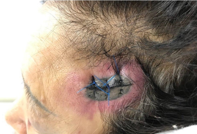
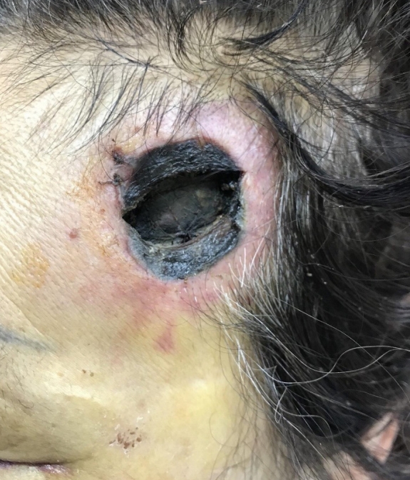
A 63-year-old woman visited the emergency department having sustained a scalp laceration. The depth of the laceration extended to the periosteum, and local wound exploration showed a crushed subcutaneous layer. Considerable amounts of bleeding and hematoma formation were observed. Rigorous hemostasis was performed by bipolar coagulation and aspiration drainage; however, hemostasis could not be achieved. Prior to skin closure and to prevent hematoma formation, three sheets of hemostatic compresses made of ORC (Surgicel) were applied above the periosteum and left in place. Five days following the procedure, overt necrosis was observed on both sides, drawing the hemostatic compresses to the posterior edge (Fig. 1). Full-thickness necrosis involving the epidermis up to the periosteal layer was observed in the affected region (Fig. 2).
ORC has been used as an absorbable hemostat since World War II [4]. ORC is a sterile bioabsorbable thrombogenic agent used adjunctively in surgical procedures to assist in the control of capillary, venous, and small arterial hemorrhage when ligation or other conventional methods of control are impractical or ineffective. One of the most commonly used ORC is Surgicel. In neurosurgery, instructions for ORC use advise against leaving them in situ in the skull or spine following hemostasis. Indeed, following tumor resection, Surgicel left in situ can induce a granulomatous inflammatory reaction to the foreign body or a chronic, persistent reaction, lasting more than 12 weeks following the procedure, creating a false impression of recurrence upon medical imaging [5]. Elsewhere, ORC use is likely to lead to paralysis or nerve damage when used around the spinal cord or to blindness in case of use in contact with the optic chiasm. Warnings in the packaging describe these risks, stating “by inflating [it], Surgicel can exert an undesirable pressure” that induces nerve injury [5].
In stomatology, leaving ORC under the mucosa has not been reported to induce similar cases of overlying necrosis [6]. In plastic surgery, Skoog used ORC to induce bone formation in the slits palatines, while other authors have demonstrated adverse effects, warning of the potential risks of cyst formation and interference with bone healing [7,8]. Erol has fabricated ORC wrapped with crushed cartilage into a “modeling clay” and used it as a nasal bridge graft for rhinoplasties performed for aesthetic reasons. The author did not report any complications following the use of ORC under the thin skin of the nasal bridge [6]. Oxidized cellulose of vegetable origin, has two active ingredients: soluble uronic acid, which is absorbed within 6 to 18 hours, and a fibrous component, which is phagocytosed by macrophages 48 hours following implantation [9,10]. Its biodegradability leads to a lowering of local pH to a range of 2.5 to 3.0 resulting in a chemical action with bactericidal activity; however, the lower pH can also compromise dermal blood flow [6]. Necrosis of muscle fibers can be observed at ORC implantation sites, likely due to their pH-lowering effects [6,10]. This may explain, in part, how tissue necrosis with a “punch” lesion occurred in this clinical case, where the hemostatic compresses were drawn to the posterior edge of the wound. The mechanical action induced by swelling of oxidized cellulose occurs due to its absorbency (up to 10 times its weight) and can also explain the resulting necrosis. We suggest that this effect was caused by separation of the tissue from the nutrient substrate, in addition to local chemical reactions. It is very likely that the combination of high concentrations of ORC in multiple sheets, left in place following the procedure, induced separation of the overlying tissue from the blood supply that was already rendered fragile by crush injury.
In conclusion, this clinical case raises concerns about the possibility of tissue necrosis following the use of ORC if left in place. Although scalp tissue is rich vascularly, separation of the bleeding skin surface, crushed skin, hematoma, and use of ORC, even in small quantities and without inducing paresthesia, can cause cutaneous pain as well as necrosis. ORC should be removed immediately once hemostasis is achieved, especially in the case of crushing injuries.